+1 (314) 370-6046 or
Contact Us - Argentina
- Australia
- Austria
- Bahrain
- Belgium
- Brazil
- Bulgaria
- Cameroon
- Canada
- Chile
- China
- Colombia
- Croatia
- Cyprus
- Czech Republic
- Denmark
- Ecuador
- Egypt
- Estonia
- Finland
- France
- Germany
- Greece
- Hong Kong
- Hungary
- Iceland
- India
- Indonesia
- Iran
- Ireland
- Israel
- Italy
- Japan
- Kazakhstan
- Kuwait
- Latvia
- Lithuania
- Luxembourg
- Macedonia
- Malaysia
- Malta
- Mexico
- Monaco
- Morocco
- Netherlands
- New Zealand
- Nigeria
- Norway
- Peru
- Philippines
- Poland
- Portugal
- Qatar
- Romania
- Russia
- Saudi Arabia
- Serbia
- Singapore
- Slovakia
- Slovenia
- South Africa
- South Korea
- Spain
- Sri Lanka
- Sweden
- Switzerland
- Taiwan
- Thailand
- Turkey
- Ukraine
- UAE
- United Kingdom
- United States
- Venezuela
- Vietnam

![Immunohistochemistry - Anti-beta Amyloid Antibody [AB9] (A85418) - Antibodies.com](https://cdn.antibodies.com/image/catalog/85/A85418_1.jpg?profile=product_top)
![Immunohistochemistry - Anti-beta Amyloid Antibody [AB9] (A85418) - Antibodies.com](https://cdn.antibodies.com/image/catalog/85/A85418_2.jpg?profile=product_top)
![Immunofluorescence - Anti-beta Amyloid Antibody [AB9] (A85418) - Antibodies.com](https://cdn.antibodies.com/image/catalog/85/A85418_3.jpg?profile=product_top)
![Western Blot - Anti-beta Amyloid Antibody [AB9] (A85418) - Antibodies.com](https://cdn.antibodies.com/image/catalog/85/A85418_4.jpg?profile=product_top)
![Western Blot - Anti-beta Amyloid Antibody [AB9] (A85418) - Antibodies.com](https://cdn.antibodies.com/image/catalog/85/A85418_5.jpg?profile=product_top)
![Immunohistochemistry - Anti-beta Amyloid Antibody [AB9] (A85418) - Antibodies.com](https://cdn.antibodies.com/image/catalog/85/A85418_1.jpg?profile=product_top_thumb)
![Immunohistochemistry - Anti-beta Amyloid Antibody [AB9] (A85418) - Antibodies.com](https://cdn.antibodies.com/image/catalog/85/A85418_2.jpg?profile=product_top_thumb)
![Immunofluorescence - Anti-beta Amyloid Antibody [AB9] (A85418) - Antibodies.com](https://cdn.antibodies.com/image/catalog/85/A85418_3.jpg?profile=product_top_thumb)
![Western Blot - Anti-beta Amyloid Antibody [AB9] (A85418) - Antibodies.com](https://cdn.antibodies.com/image/catalog/85/A85418_4.jpg?profile=product_top_thumb)
![Western Blot - Anti-beta Amyloid Antibody [AB9] (A85418) - Antibodies.com](https://cdn.antibodies.com/image/catalog/85/A85418_5.jpg?profile=product_top_thumb)
![Immunohistochemistry - Anti-beta Amyloid Antibody [AB9] (A85418) - Antibodies.com](https://cdn.antibodies.com/image/catalog/85/A85418_1.jpg?profile=product_image)
![Immunohistochemistry - Anti-beta Amyloid Antibody [AB9] (A85418) - Antibodies.com](https://cdn.antibodies.com/image/catalog/85/A85418_2.jpg?profile=product_image)
![Immunofluorescence - Anti-beta Amyloid Antibody [AB9] (A85418) - Antibodies.com](https://cdn.antibodies.com/image/catalog/85/A85418_3.jpg?profile=product_image)
![Western Blot - Anti-beta Amyloid Antibody [AB9] (A85418) - Antibodies.com](https://cdn.antibodies.com/image/catalog/85/A85418_4.jpg?profile=product_image)
![Western Blot - Anti-beta Amyloid Antibody [AB9] (A85418) - Antibodies.com](https://cdn.antibodies.com/image/catalog/85/A85418_5.jpg?profile=product_image)

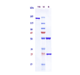
![SDS-PAGE - Anti-beta Amyloid Antibody [Research Grade Biosimilar] - Low endotoxin, Azide free (A323895) - Antibodies.com](https://cdn.antibodies.com/image/catalog/323/A323895_1.jpg?profile=product_alternative)
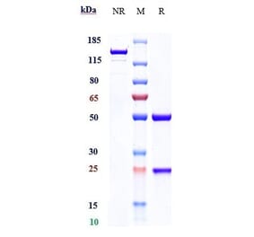
![SDS-PAGE - Anti-beta Amyloid Antibody [Research Grade Biosimilar] - Low endotoxin, Azide free (A323897) - Antibodies.com](https://cdn.antibodies.com/image/catalog/323/A323897_1.jpg?profile=product_alternative)
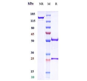
![SDS-PAGE - Anti-beta Amyloid Antibody [CNTO 2125] - Low endotoxin, Azide free (A323900) - Antibodies.com](https://cdn.antibodies.com/image/catalog/323/A323900_1.jpg?profile=product_alternative)
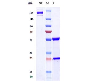
![SDS-PAGE - Anti-beta Amyloid Antibody [Research Grade Biosimilar] - Low endotoxin, Azide free (A323892) - Antibodies.com](https://cdn.antibodies.com/image/catalog/323/A323892_1.jpg?profile=product_alternative)

![SDS-PAGE - Anti-beta Amyloid Antibody [Research Grade Biosimilar] - Low endotoxin, Azide free (A323893) - Antibodies.com](https://cdn.antibodies.com/image/catalog/323/A323893_1.jpg?profile=product_alternative)
![SDS-PAGE - Anti-beta Amyloid Antibody [APP/3345] (A248989) - Antibodies.com](https://cdn.antibodies.com/image/catalog/248/A248989_1.jpg?profile=product_alternative)
![SDS-PAGE - Anti-beta Amyloid Antibody [APP/3345] - BSA and Azide free (A252169) - Antibodies.com](https://cdn.antibodies.com/image/catalog/252/A252169_1.jpg?profile=product_alternative)
![SDS-PAGE - Anti-beta Amyloid Antibody [DLX212] - Low endotoxin, Azide free (A323899) - Antibodies.com](https://cdn.antibodies.com/image/catalog/323/A323899_1.jpg?profile=product_alternative)

