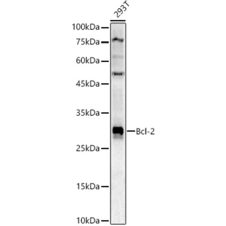
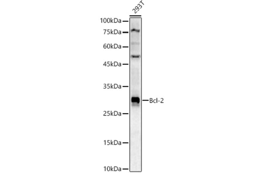
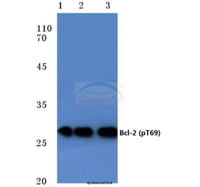
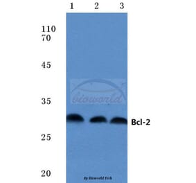
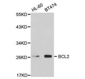
Western blot analysis of 293T, using Anti-Bcl-2 Antibody (A12540) at 1:900 dilution. The secondary antibody was Goat Anti-Rabbit IgG H&L Antibody (HRP) at 1:10,000 dilution. Lysates/proteins were present at 25µg per lane. The blocking buffer used was 3% non-fat dry milk in TBST. Detection was with a ECL Basic Kit. Exposure time: 90s.
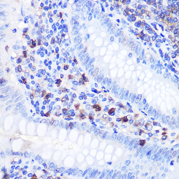
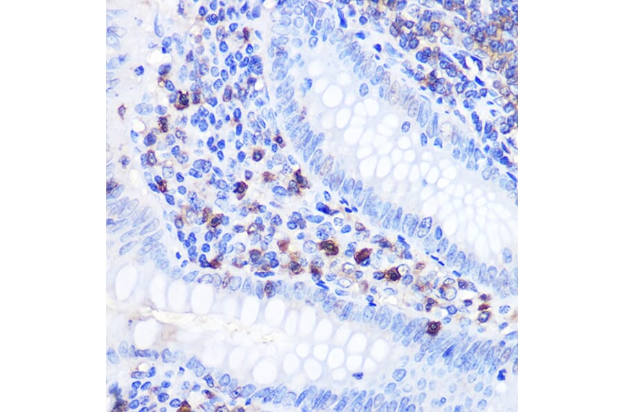
Immunohistochemistry analysis of paraffin-embedded human appendix tissue using Anti-Bcl-2 Antibody (A12540) at a dilution of 1:100 (40x lens). Perform microwave antigen retrieval with 10 mM PBS buffer pH 7.2 before commencing with IHC staining protocol.
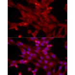
Immunofluorescence analysis of NIH/3T3 cells using Anti-Bcl-2 Antibody (A12540) at a dilution of 1:50 (40x lens). DAPI was used to stain the cell nuclei (blue).
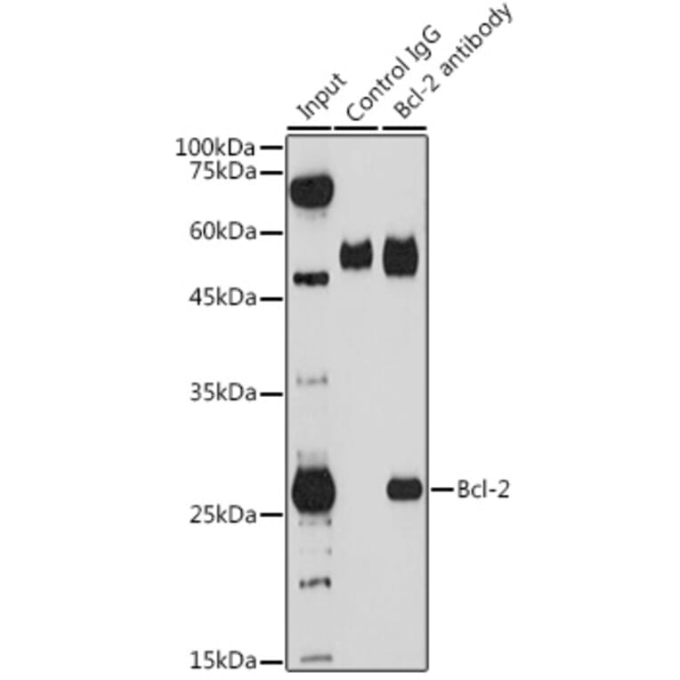
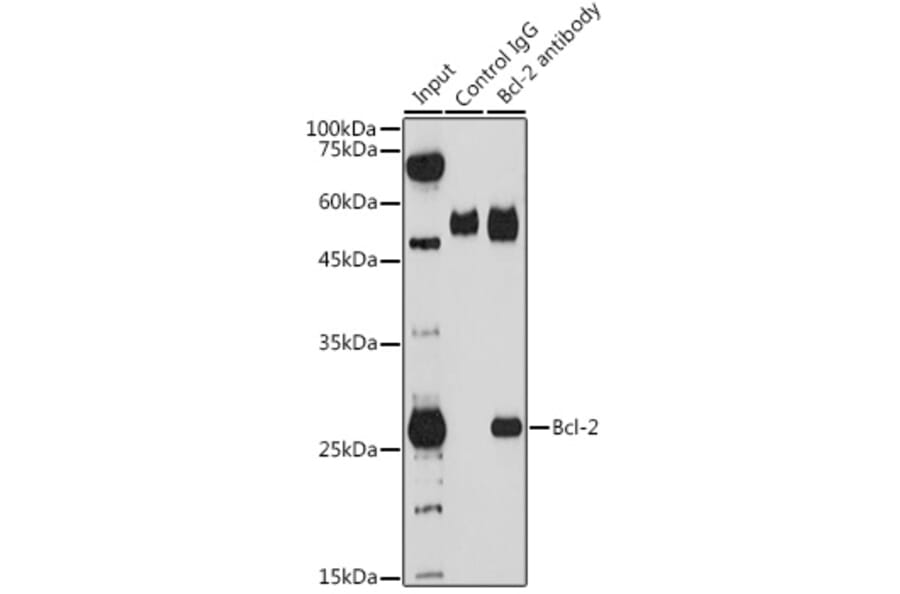
Immunoprecipitation analysis of 200µg extracts of THP-1 cells using 3µg of Anti-Bcl-2 Antibody (A12540). This Western blot was performed on the immunoprecipitate using Anti-Bcl-2 Antibody (A12540) at a dilution of 1:1000.
Publishing research using Anti-Bcl-2 Antibody (A12540)? Please let us know so that we can list the citation on this page.


![Western Blot - Anti-Bcl-2 Antibody [ARC0173] (A306910) - Antibodies.com](https://cdn.antibodies.com/image/catalog/306/A306910_1.jpg?profile=product_alternative)
![Immunohistochemistry - Anti-Bcl-2 Antibody [100/D5+124] (A249868) - Antibodies.com](https://cdn.antibodies.com/image/catalog/249/A249869_1.jpg?profile=product_alternative)
![Immunohistochemistry - Anti-Bcl-2 Antibody [100/D5] (A249863) - Antibodies.com](https://cdn.antibodies.com/image/catalog/249/A249864_1.jpg?profile=product_alternative)
![Immunohistochemistry - Anti-Bcl-2 Antibody [100/D5] - BSA and Azide free (A253043) - Antibodies.com](https://cdn.antibodies.com/image/catalog/253/A253044_1.jpg?profile=product_alternative)
![Immunohistochemistry - Anti-Bcl-2 Antibody [rBCL2/782] (A249862) - Antibodies.com](https://cdn.antibodies.com/image/catalog/249/A249862_1.jpg?profile=product_alternative)
![Immunohistochemistry - Anti-Bcl-2 Antibody [100/D5+124] - BSA and Azide free (A253048) - Antibodies.com](https://cdn.antibodies.com/image/catalog/253/A253049_1.jpg?profile=product_alternative)
![Immunohistochemistry - Anti-Bcl-2 Antibody [rBCL2/782] - BSA and Azide free (A253042) - Antibodies.com](https://cdn.antibodies.com/image/catalog/253/A253042_1.jpg?profile=product_alternative)
![Immunohistochemistry - Anti-Bcl-2 Antibody [rBCL2/796] - BSA and Azide free (A253053) - Antibodies.com](https://cdn.antibodies.com/image/catalog/253/A253053_1.jpg?profile=product_alternative)
![Immunohistochemistry - Anti-Bcl-2 Antibody [8C8] (A249865) - Antibodies.com](https://cdn.antibodies.com/image/catalog/249/A249866_1.jpg?profile=product_alternative)