Mouse monoclonal [3F11] antibody to Aurora B Kinase.
Specificity
This antibody is specific for Aurora B and does not cross-react with Aurora A and Aurora C. This antibody can be used to identify dividing or soon to be dividing cells and the antibody is also an excellent marker of midbodies both during and after cell division.
Applications
WB, ICC/IF, IHC
Dilutions
WB: 1:1,000, ICC/IF: 1:1,000, IHC: 1:1,000
Reactivity
Human, Horse, Bovine, Rat, Mouse
Immunogen
Recombinant full-length human Aurora B protein, expressed in and purified from E. coli.
Host
Mouse
Clonality
Monoclonal
Clone ID
3F11
Isotype
IgG2a
Conjugate
Unconjugated
Purification
Immunogen affinity purification.
Concentration
1 mg/ml
Molecular Weight
38 kDa
Product Form
Liquid
Formulation
Supplied in Phosphate Buffered Saline with 50% Glycerol and 5mM Sodium Azide.
Storage
Shipped at 4°C. Upon delivery aliquot and store at -20°C. Avoid freeze/thaw cycles.
Immunofluorescence - Anti-Aurora B Kinase Antibody [3F11] (A85381)
High magnification confocal immunofluorescence of HeLa cells stained with Anti-Aurora B Kinase Antibody, at a dilution of 1:1,000, in green, and Anti-Vimentin Antibody (A85421 | 1:1,000, in red. Blue is the DNA stain DAPI revealing the nuclei. The Anti-Aurora B Kinase Antibody reveals aurora B localized in midbodies, midzones of dividing cells and also in the nuclei or some cells.
Immunofluorescence - Anti-Aurora B Kinase Antibody [3F11] (A85381)
HeLa cell cultures were stained with Anti-Aurora B Kinase Antibody (green). Anti-Aurora B Kinase Antibody stains midzones in anaphase and midbodies between the two daughter cells during telophase. It is therefore a useful marker of dividing cells. Cells were counterstained with Anti-Vimentin Antibody (A85421 | red). Blue is a DNA stain.
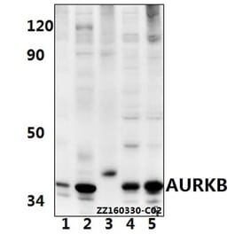
Western Blot - Anti-Aurora B Kinase Antibody [3F11] (A85381)
Western blot analysis of different cell lysates and recombinant protein solutions using Anti-Aurora B Kinase Antibody [3F11] (A85381), in green. Left: Cells were treated with 100 ng/ml of nocodazol, a microtubule depolymerizing agent which induces cells to halt at G2/M phase. The lanes contain samples of: [Lane 1] Protein standards, in red, [Lane 2] HeLa cells, [Lane 3] canine A72 cells, [Lane 4] equine NBL6 cells, and [Lane 5] mouse KR158 cells. Right: Western blot of purified full-length recombinant human Aurora A, B and C, probed with Anti-Aurora B Kinase Antibody [3F11] (A85381). The antibody binds specifically only to Aurora B and not to the closely related Aurora A and C, shown by the single band in lane B.
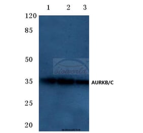
Western Blot - Anti-Aurora B Kinase Antibody [3F11] (A85381)
Left: Western blot analysis of Anti-Aurora B Kinase Antibody on lysateas from HeLa cells treated with 100ng/ml nocodazole for 18 hours. Nocodazole is a microtubule polymerization inhibitor which induces cells to halt at the G2/M phase and also induces Aurora B expression. The Anti-Aurora B Kinase Antibody binds strongly to the aurora B band at 38 kDa. Right: Blots of recombinant human Aurora A, B and C were probed with Anti-Aurora B Kinase Antibody. This antibody binds specifically only to aurora B and not to the closely related aurora A and C. This is also consistent with the immunocytochemical staining of midzones and midbodies on HeLa cells.
Publishing research using Anti-Aurora B Kinase Antibody [3F11] (A85381)? Please let us know so that we can list the citation on this page.
Alternative products to Anti-Aurora B Kinase Antibody [3F11] (A85381)


![Immunofluorescence - Anti-Aurora B Kinase Antibody [3F11] (A85381) - Antibodies.com](https://cdn.antibodies.com/image/catalog/85/A85381_1.jpg?profile=product_top)
![Immunofluorescence - Anti-Aurora B Kinase Antibody [3F11] (A85381) - Antibodies.com](https://cdn.antibodies.com/image/catalog/85/A85381_2.jpg?profile=product_top)
![Western Blot - Anti-Aurora B Kinase Antibody [3F11] (A85381) - Antibodies.com](https://cdn.antibodies.com/image/catalog/85/A85381_3.jpg?profile=product_top)
![Western Blot - Anti-Aurora B Kinase Antibody [3F11] (A85381) - Antibodies.com](https://cdn.antibodies.com/image/catalog/85/A85381_5.jpg?profile=product_top)
![Immunofluorescence - Anti-Aurora B Kinase Antibody [3F11] (A85381) - Antibodies.com](https://cdn.antibodies.com/image/catalog/85/A85381_1.jpg?profile=product_top_thumb)
![Immunofluorescence - Anti-Aurora B Kinase Antibody [3F11] (A85381) - Antibodies.com](https://cdn.antibodies.com/image/catalog/85/A85381_2.jpg?profile=product_top_thumb)
![Western Blot - Anti-Aurora B Kinase Antibody [3F11] (A85381) - Antibodies.com](https://cdn.antibodies.com/image/catalog/85/A85381_3.jpg?profile=product_top_thumb)
![Western Blot - Anti-Aurora B Kinase Antibody [3F11] (A85381) - Antibodies.com](https://cdn.antibodies.com/image/catalog/85/A85381_5.jpg?profile=product_top_thumb)
![Immunofluorescence - Anti-Aurora B Kinase Antibody [3F11] (A85381) - Antibodies.com](https://cdn.antibodies.com/image/catalog/85/A85381_1.jpg?profile=product_image)
![Immunofluorescence - Anti-Aurora B Kinase Antibody [3F11] (A85381) - Antibodies.com](https://cdn.antibodies.com/image/catalog/85/A85381_2.jpg?profile=product_image)
![Western Blot - Anti-Aurora B Kinase Antibody [3F11] (A85381) - Antibodies.com](https://cdn.antibodies.com/image/catalog/85/A85381_3.jpg?profile=product_image)
![Western Blot - Anti-Aurora B Kinase Antibody [3F11] (A85381) - Antibodies.com](https://cdn.antibodies.com/image/catalog/85/A85381_5.jpg?profile=product_image)
![Immunofluorescence - Anti-Aurora B Kinase Antibody [6G2] (A85380) - Antibodies.com](https://cdn.antibodies.com/image/catalog/85/A85380_1.jpg?profile=product_alternative)
![Western Blot - Anti-Aurora B Antibody [ARC50905] (A306974) - Antibodies.com](https://cdn.antibodies.com/image/catalog/306/A306974_1.jpg?profile=product_alternative)
![Immunohistochemistry - Anti-Aurora B Antibody [rAURKB/1592] - BSA and Azide free (A253706) - Antibodies.com](https://cdn.antibodies.com/image/catalog/253/A253706_1.jpg?profile=product_alternative)
![Immunohistochemistry - Anti-Aurora B Antibody [rAURKB/1592] (A250526) - Antibodies.com](https://cdn.antibodies.com/image/catalog/250/A250526_1.jpg?profile=product_alternative)
![Immunohistochemistry - Anti-Aurora B Antibody [AURKB/1592] (A250522) - Antibodies.com](https://cdn.antibodies.com/image/catalog/250/A250523_1.jpg?profile=product_alternative)
![Immunohistochemistry - Anti-Aurora B Antibody [AURKB/1592] - BSA and Azide free (A253702) - Antibodies.com](https://cdn.antibodies.com/image/catalog/253/A253703_1.jpg?profile=product_alternative)
![Immunohistochemistry - Anti-Aurora B Antibody [RM278] (A121378) - Antibodies.com](https://cdn.antibodies.com/image/catalog/121/A121498_1.png?profile=product_alternative)
![Immunohistochemistry - Anti-Aurora B Antibody [AURKB/1593] (A250524) - Antibodies.com](https://cdn.antibodies.com/image/catalog/250/A250524_1.jpg?profile=product_alternative)
![Immunohistochemistry - Anti-Aurora B Antibody [AURKB/1593] - BSA and Azide free (A253704) - Antibodies.com](https://cdn.antibodies.com/image/catalog/253/A253704_1.jpg?profile=product_alternative)