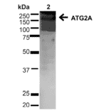
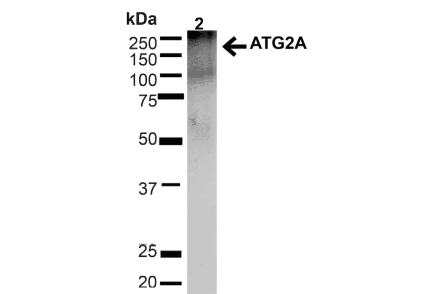
Western blot analysis of human Cervical cancer cell line (HeLa) lysate showing detection of ~212.9kDa ATG2A protein using Anti-ATG2A Antibody (A304941) at 1:1,000 for 1 hour at room temperature. Lane 1: MW Ladder. Lane 2: human HeLa (20 µg). Load: 20 µg. Block: 5% milk + TBST for 1 hour at room temperature. The secondary antibody used was Goat Anti-Rabbit: HRP at 1:2000 for 1 hour at room temperature. Color Development: TMB solution for 12 min at room temperature. Predicted/Observed Size: ~212.9kDa.
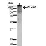
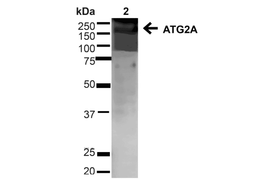
Western blot analysis of mouse brain showing detection of ~212.9kDa ATG2A protein using Anti-ATG2A Antibody (A304941) at 1:1,000 for 1 hour at room temperature. Lane 1: MW Ladder. Lane 2: mouse brain (20 µg). Load: 20 µg. Block: 5% milk + TBST for 1 hour at room temperature. The secondary antibody used was Goat Anti-Rabbit: HRP at 1:2000 for 1 hour at room temperature. Color Development: TMB solution for 12 min at room temperature. Predicted/Observed Size: ~212.9kDa.
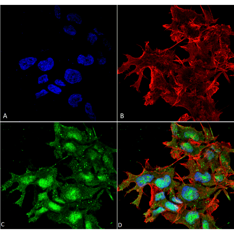
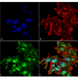
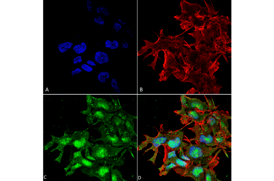
Immunocytochemistry/Immunofluorescence analysis of human neuroblastoma cell line (SK-N-BE, fixed in 4% formaldehyde for 15 min at room temperature, using Anti-ATG2A Antibody (A304941), at 1:100 for 60 minutes at room temperature. The secondary antibody used was Goat Anti-Rabbit ATTO 488 at 1:100 for 60 minutes at room temperature. Counterstain: Phalloidin Texas Red F-Actin stain; DAPI (blue) nuclear stain at 1:1000, 1:5,000 for 60min room temperature, 5min room temperature. Localization: Nucleus, Cytoplasm. Magnification: 60X.(A) DAPI (blue) nuclear stain (B) Phalloidin Texas Red F-Actin stain (C) ATG2A Antibody (D) Composite.
Publishing research using Anti-ATG2A Antibody (A304941)? Please let us know so that we can list the citation on this page.
Alternative products to Anti-ATG2A Antibody (A304941)