Recombinant full-length human alpha Synuclein protein, region 61-95 corresponding to the central “non-amyloid beta component of Alzheimer’s disease amyloid”, expressed in and purified from E. coli.
Host
Chicken
Clonality
Polyclonal
Isotype
IgY
Conjugate
Unconjugated
Purification
Immunogen affinity purification.
Concentration
1 mg/ml
Molecular Weight
15 kDa (by SDS-PAGE)
Product Form
Liquid
Formulation
Supplied in Phosphate Buffered Saline with 50% Glycerol and 5mM Sodium Azide.
Storage
Shipped at 4°C. Store at +4°C. Do not freeze!
Synonyms
Alpha-synuclein, NACP, Non-A beta component of AD amyloid, Non-A4 component of amyloid precursor, PARK1, SNCA
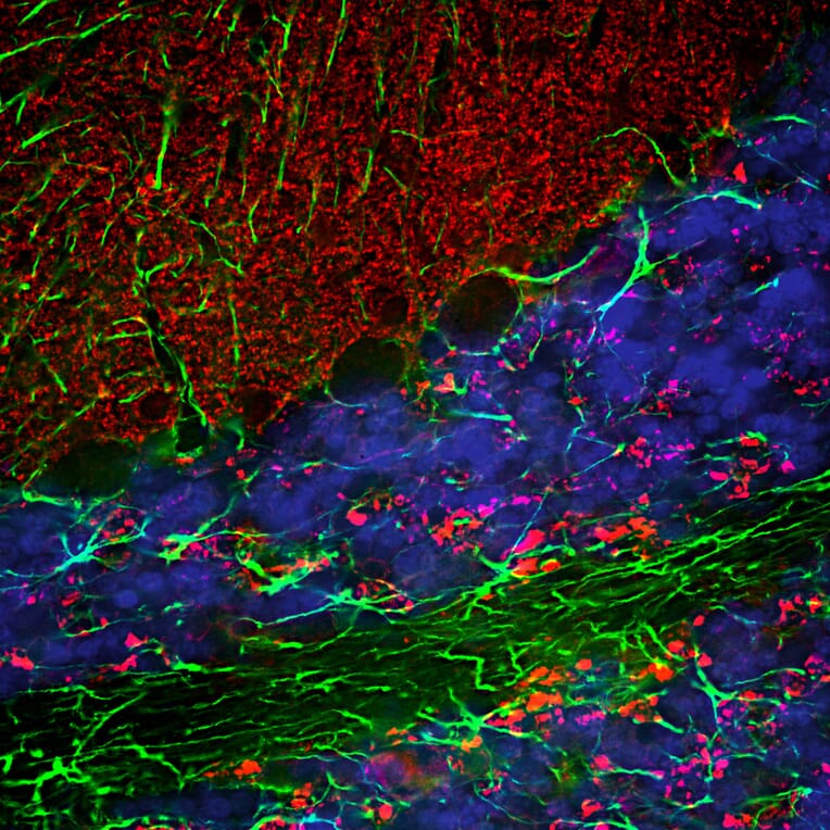
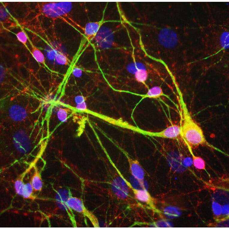
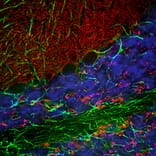
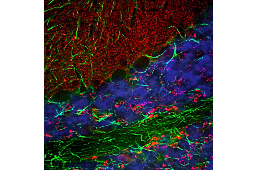
Immunofluorescent analysis of a section of rat cerebellum stained with Anti-Alpha-synuclein Antibody (A85289), dilution 1:3,000, in red, and co-stained with Anti-GFAP Antibody (A85419), dilution 1:5,000, in green. The blue is DAPI staining of nuclear DNA. Following transcardial perfusion of the rat with 4% paraformaldehyde, the brain was post fixed for 24 hours, cut to 45 µm, and free-floating sections were stained with above antibodies. The alpha-synuclein protein is concentrated in presynaptic regions in the granular and molecular layers, while the GFAP antibody stains the network of Bergmann and astroglial cells.
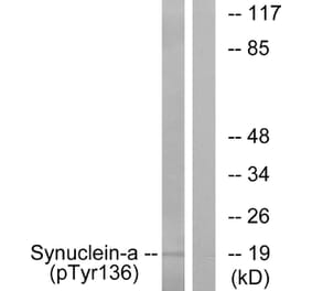
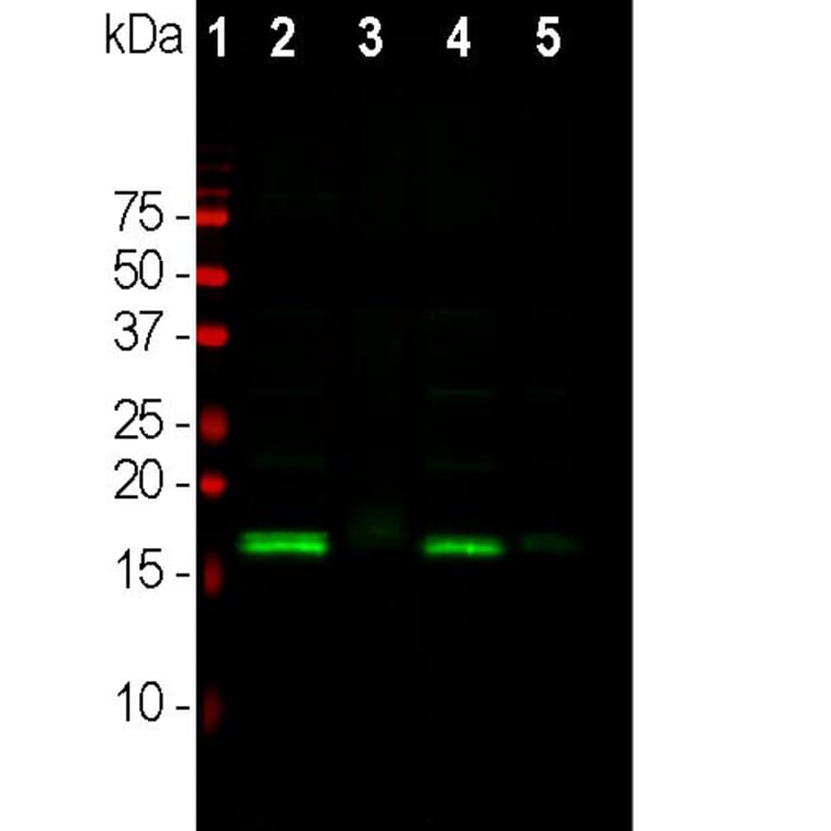
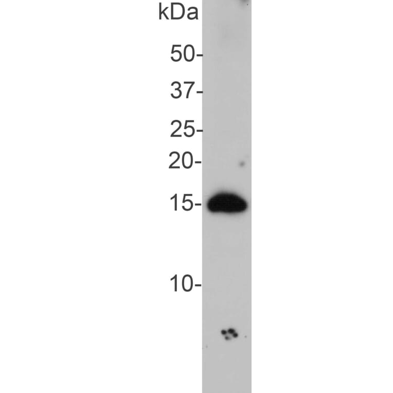
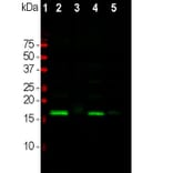
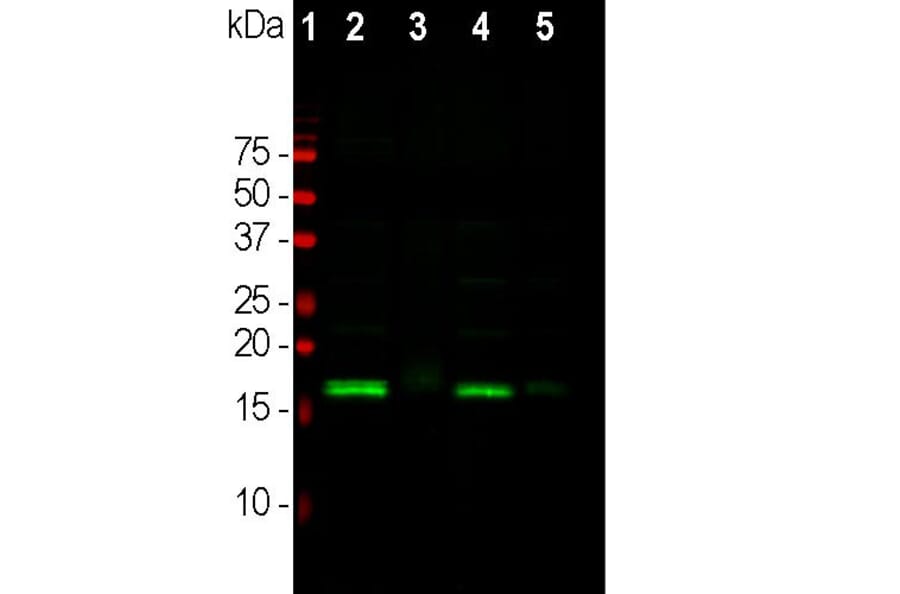
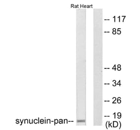
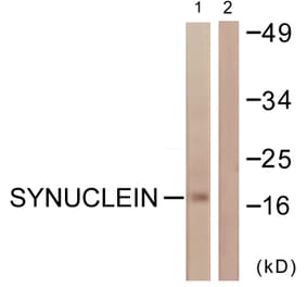
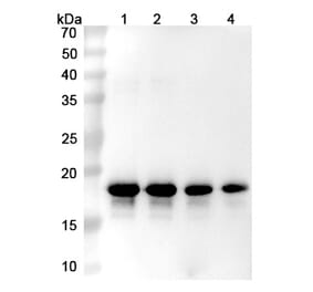
Western Blot - Anti-alpha Synuclein Antibody (A85289)
Western blot analysis of different tissue lysates using Anti-Alpha-synuclein Antibody (A85289), dilution 1:2,000 in green. The lanes contain: [Lane 1] protein standard (red), [Lane 2] rat brain, [Lane 3] rat spinal cord, [Lane 4] mouse brain, and [Lane 5] mouse spinal cord. The strong band at about 15 kDa corresponds to the alpha-synuclein protein in brain extracts, which are rich in synapses, while a weaker band is seen in spinal cord extracts where synapses are a more minor component.
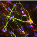
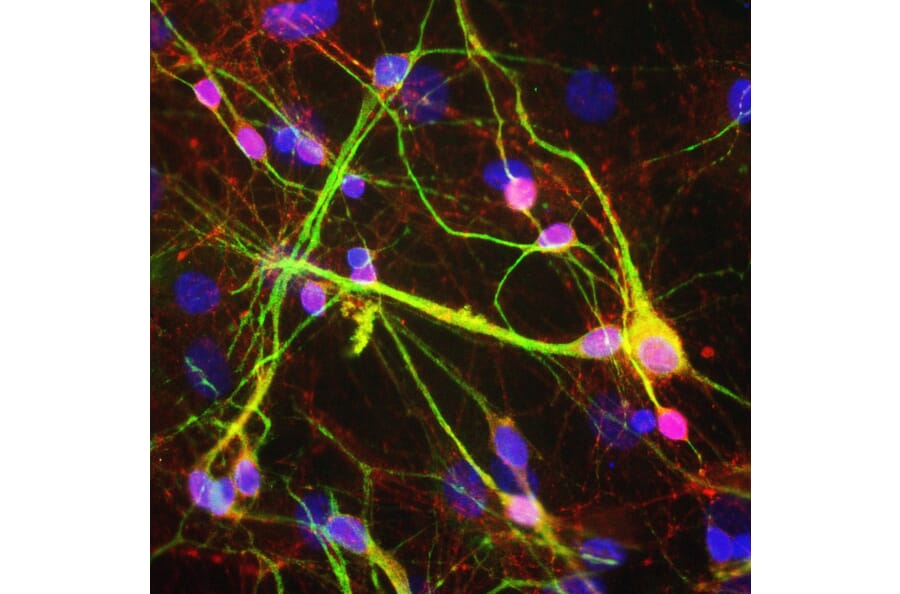
Mixed rat neuron-glial cultures stained with Anti-Alpha-Synuclein Antibody (red) and Anti-MAP2 Antibody (green). The alpha-synuclein antibody stains vesicular structures - the perikarya and processes of the neurons in this image. Note that some of the neuronal perikarya contain much more alpha-synuclein than others. The blue channel shows the localization of DNA.
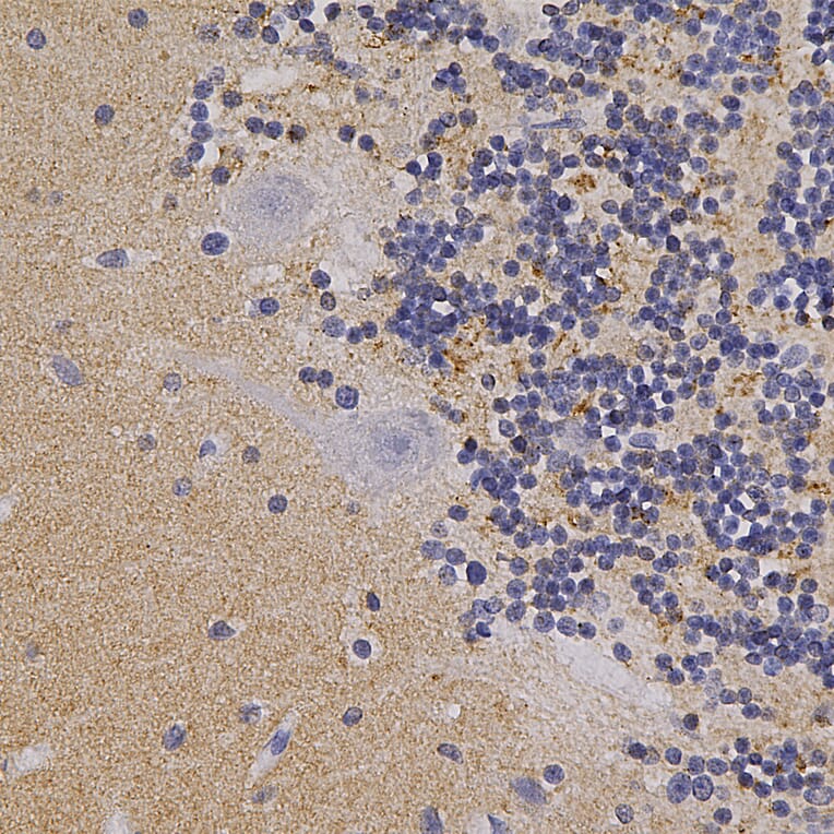
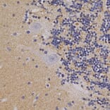
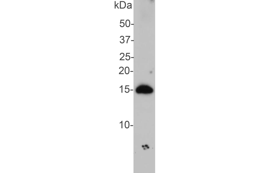
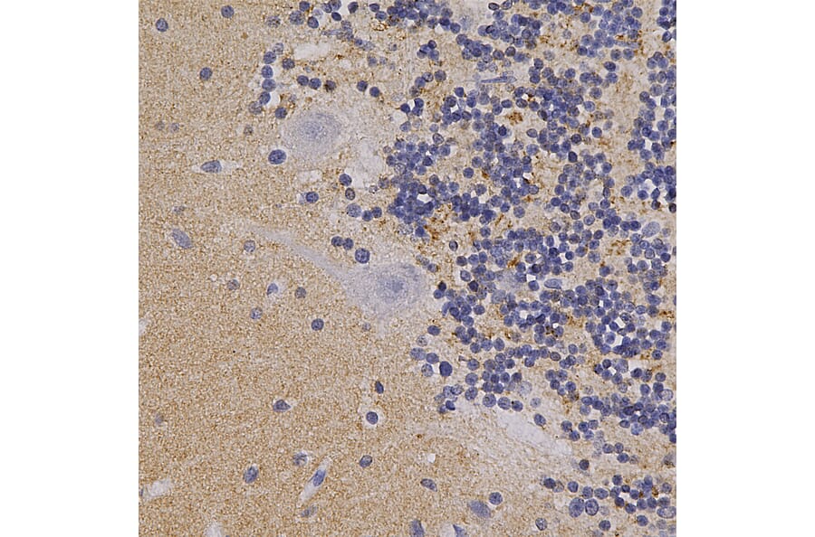
Immunohistochemistry analysis of a formalin fixed paraffin embedded human cerebellum section with Anti-alpha Synuclein Antibody (A85289) at a dilution of 1:5,000 detected in DAB (brown) following the ABC method. Counterstained with Hematoxylin (blue). The a-synuclein protein is concentrated in synapses, in particular highlighting the glomeruli synapses in the granule layer and those on Purkinje dendrites in the molecular layer.
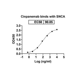
Humanized monoclonal antibody to alpha Synuclein Research grade Cinpanemab biosimilar for ELISA, Flow Cytometry, Functional Studies and in vivo Research.
Humanized monoclonal antibody to alpha Synuclein Research grade Prasinezumab biosimilar for ELISA, Flow Cytometry, Functional Studies and in vivo Research.
Human monoclonal antibody to alpha Synuclein Research grade Anti-Human Alpha-synuclein [Lu AF82422] for ELISA, Flow Cytometry, Functional Studies and in vivo Research.


![Immunofluorescence - Anti-alpha Synuclein Antibody [2A7] (A85290) - Antibodies.com](https://cdn.antibodies.com/image/catalog/85/A85290_1.jpg?profile=product_alternative)
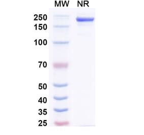
![SDS-PAGE - Anti-alpha Synuclein Antibody [MEDI1341] Biosimilar - BSA and Azide free (A339878) - Antibodies.com](https://cdn.antibodies.com/image/catalog/339/A339878_1.jpg?profile=product_alternative)
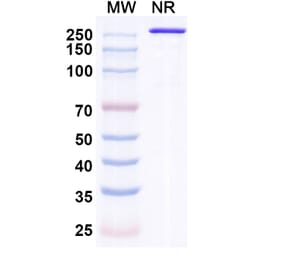
![SDS-PAGE - Anti-alpha Synuclein Antibody [Lu AF82422] Biosimilar - BSA and Azide free (A339330) - Antibodies.com](https://cdn.antibodies.com/image/catalog/339/A339330_1.jpg?profile=product_alternative)