Mouse monoclonal [2A7] antibody to alpha Synuclein.
Specificity
This antibody specifically recognizes alpha Synuclein and also binds human alpha Synuclein containing either the A30P or the A53T Parkinson’s associated mutations. This antibody does not cross-react with beta Synuclein or gamma Synuclein. The epitope is in the region 61-95; which corresponds to the “Non-amyloid beta component of Alzheimer’s disease amyloid”.
Applications
WB, ICC/IF, IHC
Dilutions
WB: 1:1,000, ICC/IF: 1:1,000
Reactivity
Human, Bovine, Porcine, Rat, Mouse
Immunogen
Recombinant full-length human alpha Synuclein, expressed in and purified from E. coli.
Host
Mouse
Clonality
Monoclonal
Clone ID
2A7
Isotype
IgG1
Light Chains
kappa
Conjugate
Unconjugated
Purification
Immunogen affinity purification.
Concentration
1 mg/ml
Molecular Weight
~15 kDa
Product Form
Liquid
Formulation
Supplied in Phosphate Buffered Saline with 50% Glycerol and 5mM Sodium Azide.
Storage
Shipped at 4°C. Upon delivery aliquot and store at -20°C. Avoid freeze/thaw cycles.
Synonyms
Alpha-synuclein, NACP, Non-A beta component of AD amyloid, Non-A4 component of amyloid precursor, PARK1, SNCA
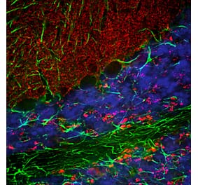
Immunofluorescent analysis of rat cerebellum section co-stained with Anti-a-Synuclein Antibody, at a dilution of 1:1,000, in red, and Anti-GFAP Antibody (A85419 | 1:5,000, in green. The blue is DAPI staining of nuclear DNA. Following transcardial perfusion of rat with 4% paraformaldehyde, brain was post fixed for 24 hours, cut to 45µM, and free-floating sections were stained with the above antibodies. The a-synuclein protein is concentrated in synaptic regions, while the Anti-GFAP Antibody stains the filamentous cytoskeleton of Bergmann glia and astrocytic cells.
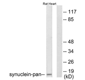
Western Blot - Anti-alpha Synuclein Antibody [2A7] (A85290)
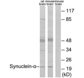
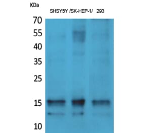
Western blot analysis of tissue lysates using Anti-a-Synuclein Antibody, at a dilution of 1:1,000, in green,: [Lane 1] protein standard in red, [Lane 2] whole rat brain lysate, [Lane 3] rat spinal cord lysate. Strong band at about 15kDa corresponds to a-synuclein protein.
Mixed neuron-glial cultures stained with Anti-a-Synuclein Antibody (red) and Anti-MAP2 Antibody (A85363 | green). The Anti-a-Synuclein Antibody stains vesicular structures - the perikarya and processes of the neurons in this image. Note that some of the neuronal perikarya contain much more alpha-synuclein than others. The blue channel shows the localization of DNA.
Immunofluorescent analysis of rat olfactory bulb section stained with Anti-MeCP2 Antibody (A104322), at a dilution of 1:2,000, in red, and co-stained with Anti-alpha Synuclein Antibody [2A7] (A85290), at a dilution of 1:1,000, in green. The blue is DAPI staining of nuclear DNA. Following transcardial perfusion of rat with 4% paraformaldehyde, brain was post fixed for 24 hours, cut to 45µM, and free-floating sections were stained with the above antibodies. Anti-MeCP2 Antibody (A104322) specifically labels the nuclei of neuronal cells while Anti-alpha Synuclein Antibody [2A7] (A85290) reveals alpha Synuclein protein concentrated in presynaptic regions.
Dot blot analysis of various truncated and mutant construct applied to PVDF membranes in about of 400ng of each protein. These were the first 60 amino acids of Anti-alpha Synuclein Antibody [2A7] (A85290), amino acids 61 to 140 (61-140), full length but incorporating the A30P and A53T mutations seen associated with familial forms of Parkinson’s disease (A30P/A53T), full length but with the central NAC region of amino acids 61-95 missing (d61-95) and full length alpha synuclein. The epitopes for Anti-alpha Synuclein Antibody [2A7] (A85290) are clearly in the NAC region from 61-95 within amino acids 61-140, but do not appear to bind amino acids 1-60, suggesting that this region has low immunogenicity.
Immunofluorescent analysis of rat hippocampus section costained with Anti-alpha Synuclein Antibody [2A7] (A85290) at a dilution of 1:1,000 (green) and Anti-MeCP2 Antibody (A104322) at a dilution of 1:2,000 (red). Nuclei were stained with DAPI (blue). Following transcardial perfusion of rat with 4% paraformaldehyde, brain was post fixed for 1 hour, cut to 45 µM, and free-floating sections were stained with above antibodies. The a-synuclein protein is concentrated in synaptic regions, and the MeCP2 antibody stains the nuclei of neuronal cells.
Immunofluorescent analysis of rat olfactory bulb section costained with Anti-alpha Synuclein Antibody [2A7] (A85290) at a dilution of 1:1,000 (red) and Anti-GFAP Antibody (A85419) at a dilution of 1:5,000 (green). Nuclei were stained with DAPI (blue). Following transcardial perfusion of rat with 4% paraformaldehyde, brain was post fixed for 24 hours, cut to 45µM, and free floating sections were stained with above antibodies. The a-synuclein protein is concentrated in synaptic regions, while the GFAP antibody stains the filamentous backbone of astroglial cells.
Immunohistochemistry analysis of a NBF fixed paraffin embedded human cerebellum section with Anti-alpha Synuclein Antibody [2A7] (A85290) at a dilution of 1:1,000 detected in DAB (brown) using the Vector Labs ImmPRESS method and reagents with citra buffer retrieval. Counterstained with Hematoxylin (blue). The a-synuclein antibody synaptic regions in the granular and molecular layers of the cerebellum. Note: this antibody performs well in testing with both 4% PFA and standard NBF fixed tissues.
Immunohistochemistry analysis of the cortex of a patient with Parkinson’s disease (PD) stained with Anti-alpha Synuclein Antibody [2A7] (A85290) at a dilution of 1:1,000 with horse radish peroxidase and DAB. The Lewy bodies and other typical inclusions of PD are seen in brown.
Western Blot - Anti-alpha Synuclein Antibody [2A7] (A85290)
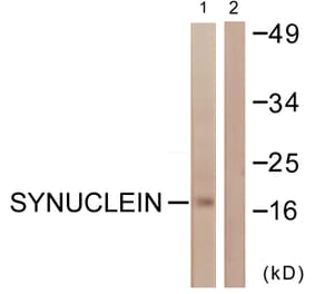
Western blot (left) and Ponceau S stained blot (right) analysis of [Lane 1] protein standard, recombinant full length human [Lane 2] alpha, [Lane 3] beta, and [Lane 4] gamma Synuclein in lanes 2, 3 and 4 respectively. Anti-alpha Synuclein Antibody [2A7] (A85290) reacts strongly with alpha Synuclein and shows no reaction with the other proteins.


![Immunofluorescence - Anti-alpha Synuclein Antibody [2A7] (A85290) - Antibodies.com](https://cdn.antibodies.com/image/catalog/85/A85290_1.jpg?profile=product_top)
![Western Blot - Anti-alpha Synuclein Antibody [2A7] (A85290) - Antibodies.com](https://cdn.antibodies.com/image/catalog/85/A85290_2.jpg?profile=product_top)
![Immunofluorescence - Anti-alpha Synuclein Antibody [2A7] (A85290) - Antibodies.com](https://cdn.antibodies.com/image/catalog/85/A85290_3.jpg?profile=product_top)
![Western Blot - Anti-alpha Synuclein Antibody [2A7] (A85290) - Antibodies.com](https://cdn.antibodies.com/image/catalog/85/A85290_5.jpg?profile=product_top)
![Immunofluorescence - Anti-alpha Synuclein Antibody [2A7] (A85290) - Antibodies.com](https://cdn.antibodies.com/image/catalog/85/A85290_6.jpg?profile=product_top)
![Dot blot - Anti-alpha Synuclein Antibody [2A7] (A85290) - Antibodies.com](https://cdn.antibodies.com/image/catalog/85/A85290_7.jpg?profile=product_top)
![Immunofluorescence - Anti-alpha Synuclein Antibody [2A7] (A85290) - Antibodies.com](https://cdn.antibodies.com/image/catalog/85/A85290_8.jpg?profile=product_top)
![Immunofluorescence - Anti-alpha Synuclein Antibody [2A7] (A85290) - Antibodies.com](https://cdn.antibodies.com/image/catalog/85/A85290_9.jpg?profile=product_top)
![Immunohistochemistry - Anti-alpha Synuclein Antibody [2A7] (A85290) - Antibodies.com](https://cdn.antibodies.com/image/catalog/85/A85290_10.jpg?profile=product_top)
![Immunohistochemistry - Anti-alpha Synuclein Antibody [2A7] (A85290) - Antibodies.com](https://cdn.antibodies.com/image/catalog/85/A85290_11.jpg?profile=product_top)
![Western Blot - Anti-alpha Synuclein Antibody [2A7] (A85290) - Antibodies.com](https://cdn.antibodies.com/image/catalog/85/A85290_12.jpg?profile=product_top)
![Immunofluorescence - Anti-alpha Synuclein Antibody [2A7] (A85290) - Antibodies.com](https://cdn.antibodies.com/image/catalog/85/A85290_1.jpg?profile=product_top_thumb)
![Western Blot - Anti-alpha Synuclein Antibody [2A7] (A85290) - Antibodies.com](https://cdn.antibodies.com/image/catalog/85/A85290_2.jpg?profile=product_top_thumb)
![Immunofluorescence - Anti-alpha Synuclein Antibody [2A7] (A85290) - Antibodies.com](https://cdn.antibodies.com/image/catalog/85/A85290_3.jpg?profile=product_top_thumb)
![Western Blot - Anti-alpha Synuclein Antibody [2A7] (A85290) - Antibodies.com](https://cdn.antibodies.com/image/catalog/85/A85290_5.jpg?profile=product_top_thumb)
![Immunofluorescence - Anti-alpha Synuclein Antibody [2A7] (A85290) - Antibodies.com](https://cdn.antibodies.com/image/catalog/85/A85290_6.jpg?profile=product_top_thumb)
![Dot blot - Anti-alpha Synuclein Antibody [2A7] (A85290) - Antibodies.com](https://cdn.antibodies.com/image/catalog/85/A85290_7.jpg?profile=product_top_thumb)
![Immunofluorescence - Anti-alpha Synuclein Antibody [2A7] (A85290) - Antibodies.com](https://cdn.antibodies.com/image/catalog/85/A85290_8.jpg?profile=product_top_thumb)
![Immunofluorescence - Anti-alpha Synuclein Antibody [2A7] (A85290) - Antibodies.com](https://cdn.antibodies.com/image/catalog/85/A85290_1.jpg?profile=product_image)
![Western Blot - Anti-alpha Synuclein Antibody [2A7] (A85290) - Antibodies.com](https://cdn.antibodies.com/image/catalog/85/A85290_2.jpg?profile=product_image)
![Immunofluorescence - Anti-alpha Synuclein Antibody [2A7] (A85290) - Antibodies.com](https://cdn.antibodies.com/image/catalog/85/A85290_3.jpg?profile=product_image)
![Western Blot - Anti-alpha Synuclein Antibody [2A7] (A85290) - Antibodies.com](https://cdn.antibodies.com/image/catalog/85/A85290_5.jpg?profile=product_image)
![Immunofluorescence - Anti-alpha Synuclein Antibody [2A7] (A85290) - Antibodies.com](https://cdn.antibodies.com/image/catalog/85/A85290_6.jpg?profile=product_image)
![Dot blot - Anti-alpha Synuclein Antibody [2A7] (A85290) - Antibodies.com](https://cdn.antibodies.com/image/catalog/85/A85290_7.jpg?profile=product_image)
![Immunofluorescence - Anti-alpha Synuclein Antibody [2A7] (A85290) - Antibodies.com](https://cdn.antibodies.com/image/catalog/85/A85290_8.jpg?profile=product_image)
![Immunofluorescence - Anti-alpha Synuclein Antibody [2A7] (A85290) - Antibodies.com](https://cdn.antibodies.com/image/catalog/85/A85290_9.jpg?profile=product_image)
![Immunohistochemistry - Anti-alpha Synuclein Antibody [2A7] (A85290) - Antibodies.com](https://cdn.antibodies.com/image/catalog/85/A85290_10.jpg?profile=product_image)
![Immunohistochemistry - Anti-alpha Synuclein Antibody [2A7] (A85290) - Antibodies.com](https://cdn.antibodies.com/image/catalog/85/A85290_11.jpg?profile=product_image)
![Western Blot - Anti-alpha Synuclein Antibody [2A7] (A85290) - Antibodies.com](https://cdn.antibodies.com/image/catalog/85/A85290_12.jpg?profile=product_image)

![Immunocytochemistry/Immunofluorescence - Anti-alpha Synuclein (phospho Ser129) Antibody [J18] (A304933) - Antibodies.com](https://cdn.antibodies.com/image/catalog/304/A304933_1.png?profile=product_alternative)
![Western Blot - Anti-alpha Synuclein Antibody [3C11] (A304959) - Antibodies.com](https://cdn.antibodies.com/image/catalog/304/A304959_1.png?profile=product_alternative)
![Immunocytochemistry/Immunofluorescence - Anti-alpha Synuclein Antibody [4F1] (A304962) - Antibodies.com](https://cdn.antibodies.com/image/catalog/304/A304962_1.png?profile=product_alternative)