Immunofluorescent analysis of cortical neuron-glial cell culture from E20 rat stained with Anti-ALDH1L1 Antibody [2E7] (A85314), dilution 1:1,000, in red, and co-stained with Anti-GFAP Antibody (A85307), dilution 1:5,000 in green. The blue is Hoechst staining of nuclear DNA. The Anti-ALDH1L1 Antibody [2E7] produces cytoplasmic staining of glial cells, while the Anti-GFAP Antibody labels the intermediate filament cytoskeleton in astrocytes and other glial cells. Some astrocytic cells express both ALDH1L1 and GFAP and therefore appear yellow.
Western Blot - Anti-ALDH1L1 Antibody [2E7] (A85314)
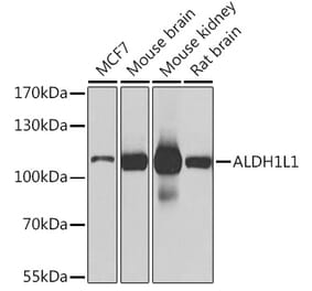
Western blot analysis of different tissue and cell lysates using Anti-ALDH1L1 Antibody [2E7] (A85314), dilution 1:5,000, in green. The lanes contain: [Lane 1] protein standard (red), rat tissue lysates: [Lane 2] heart, [Lane 3] liver, [Lane 4] kidney, [Lane 5] lung, [Lane 6] brain, and [Lane 7] spinal cord; mouse tissue lysates: [Lane 8] brain, and [Lane 9] spinal cord; cell lysates: [Lane 10] NIH-3T3, and [Lane 11] HEK293. The band at 100kDa mark corresponds to ALDH1L1 protein.
Neuron-glia cell mixed cultures stained with Anti-Aldehyde Dehydrogenase H1L1 Antibody (red) and Anti-Vimentin Antibody (A85421 | green). Blue is a DNA stain. Anti-Aldehyde Dehydrogenase H1L1 Antibody stains astrocytic cell bodies and processes. The fibroblastic cells contain only vimentin and so are green, while astrocytes contain either vimentin and ALDH1L1, so appearing golden, or predominantly ALDH1L1, in which case they appear red.
Western Blot - Anti-ALDH1L1 Antibody [2E7] (A85314)
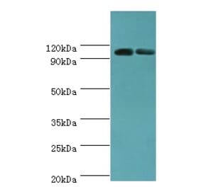
Blot of rat liver tissue homogenates blotted with Anti-Aldehyde Dehydrogenase H1L1 Antibody. Anti-Aldehyde Dehydrogenase H1L1 Antibody binding produces a strong band at ~100 kDa.
Publishing research using Anti-ALDH1L1 Antibody [2E7] (A85314)? Please let us know so that we can list the citation on this page.
Alternative products to Anti-ALDH1L1 Antibody [2E7] (A85314)


![Immunofluorescence - Anti-ALDH1L1 Antibody [2E7] (A85314) - Antibodies.com](https://cdn.antibodies.com/image/catalog/85/A85314_1.jpg?profile=product_top)
![Western Blot - Anti-ALDH1L1 Antibody [2E7] (A85314) - Antibodies.com](https://cdn.antibodies.com/image/catalog/85/A85314_2.jpg?profile=product_top)
![Immunofluorescence - Anti-ALDH1L1 Antibody [2E7] (A85314) - Antibodies.com](https://cdn.antibodies.com/image/catalog/85/A85314_3.jpg?profile=product_top)
![Western Blot - Anti-ALDH1L1 Antibody [2E7] (A85314) - Antibodies.com](https://cdn.antibodies.com/image/catalog/85/A85314_4.jpg?profile=product_top)
![Immunofluorescence - Anti-ALDH1L1 Antibody [2E7] (A85314) - Antibodies.com](https://cdn.antibodies.com/image/catalog/85/A85314_1.jpg?profile=product_top_thumb)
![Western Blot - Anti-ALDH1L1 Antibody [2E7] (A85314) - Antibodies.com](https://cdn.antibodies.com/image/catalog/85/A85314_2.jpg?profile=product_top_thumb)
![Immunofluorescence - Anti-ALDH1L1 Antibody [2E7] (A85314) - Antibodies.com](https://cdn.antibodies.com/image/catalog/85/A85314_3.jpg?profile=product_top_thumb)
![Western Blot - Anti-ALDH1L1 Antibody [2E7] (A85314) - Antibodies.com](https://cdn.antibodies.com/image/catalog/85/A85314_4.jpg?profile=product_top_thumb)
![Immunofluorescence - Anti-ALDH1L1 Antibody [2E7] (A85314) - Antibodies.com](https://cdn.antibodies.com/image/catalog/85/A85314_1.jpg?profile=product_image)
![Western Blot - Anti-ALDH1L1 Antibody [2E7] (A85314) - Antibodies.com](https://cdn.antibodies.com/image/catalog/85/A85314_2.jpg?profile=product_image)
![Immunofluorescence - Anti-ALDH1L1 Antibody [2E7] (A85314) - Antibodies.com](https://cdn.antibodies.com/image/catalog/85/A85314_3.jpg?profile=product_image)
![Western Blot - Anti-ALDH1L1 Antibody [2E7] (A85314) - Antibodies.com](https://cdn.antibodies.com/image/catalog/85/A85314_4.jpg?profile=product_image)
![Immunofluorescence - Anti-ALDH1L1 Antibody [4A12] (A85315) - Antibodies.com](https://cdn.antibodies.com/image/catalog/85/A85315_1.jpg?profile=product_alternative)