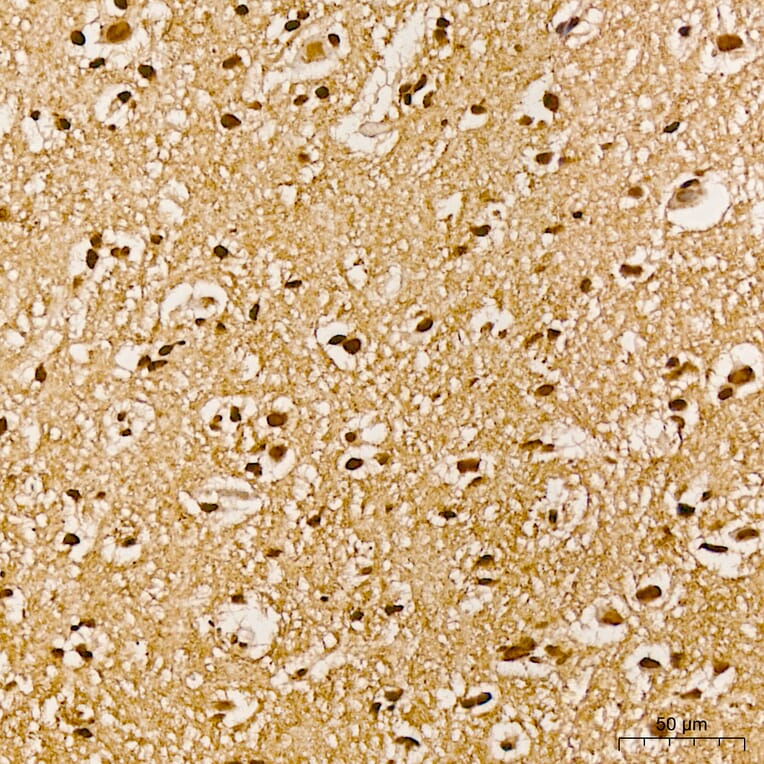
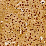
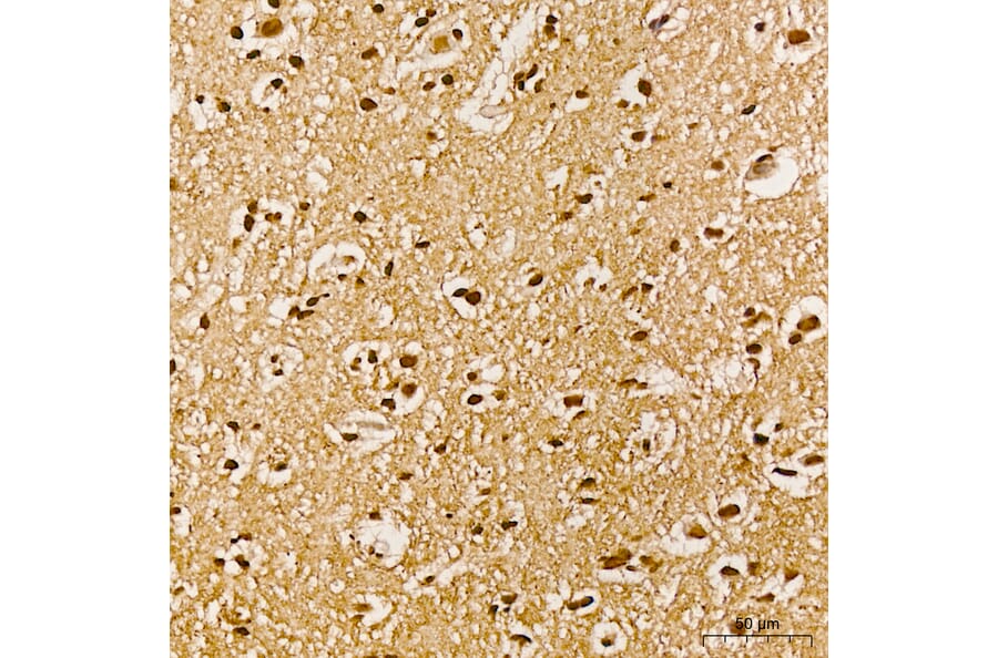
AKT1 expression in human brain tissue analyzed by immunohistochemistry. Tissue was paraffin-embedded, and antigen retrieval was achieved with 10 mM citrate buffer, pH 6.0, under high pressure. Staining was performed with Anti-AKT1 (Phospho Y315) Antibody (A16561) at a dilution of 1:100.
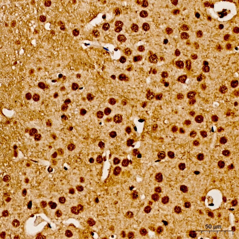
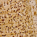
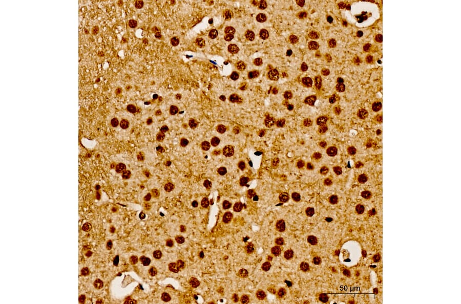
AKT1 expression in mouse brain tissue analyzed by immunohistochemistry. Tissue was paraffin-embedded, and antigen retrieval was achieved with 10 mM citrate buffer, pH 6.0, under high pressure. Staining was performed with Anti-AKT1 (Phospho Y315) Antibody (A16561) at a dilution of 1:100.
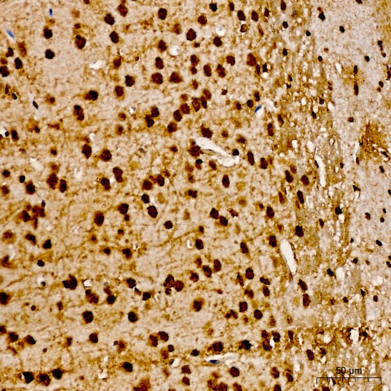
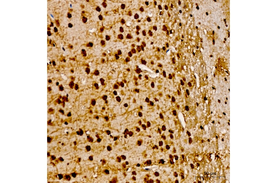
AKT1 expression in rat brain tissue analyzed by immunohistochemistry. Tissue was paraffin-embedded, and antigen retrieval was achieved with 10 mM citrate buffer, pH 6.0, under high pressure. Staining was performed with Anti-AKT1 (Phospho Y315) Antibody (A16561) at a dilution of 1:100.
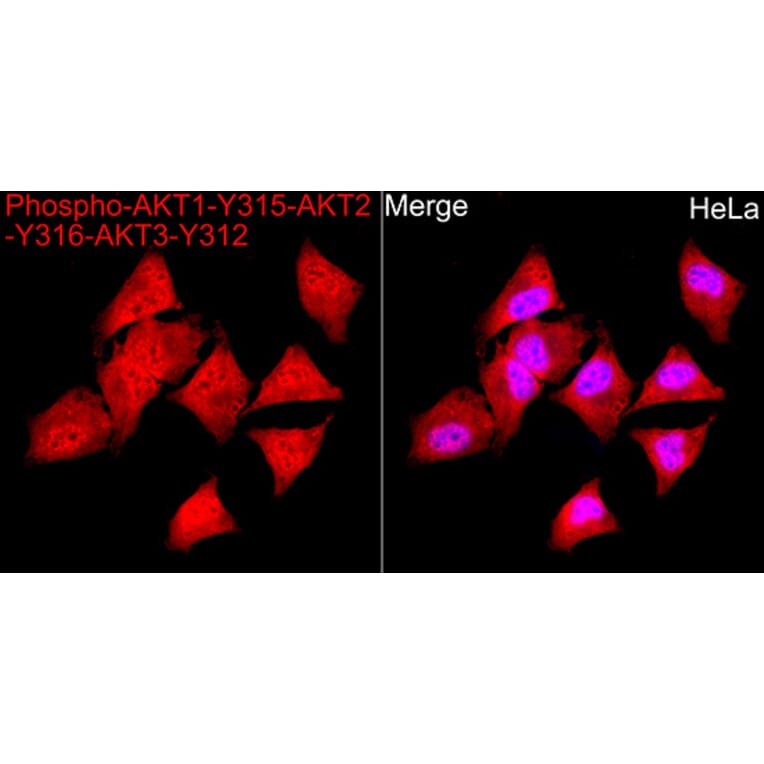
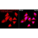
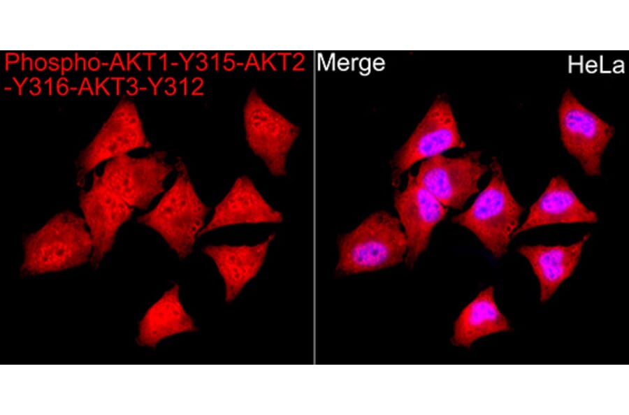
AKT1 expression in Hela cells analyzed by immunofluorescence. Staining was performed with Anti-AKT1 (Phospho Y315) Antibody (A16561) at a dilution of 1:100 followed by Cy3-conjugated Goat anti-Rabbit IgG (H+L) secondary antibody at a dilution of 1:500. Nuclei were stained with DAPI (blue).
Publishing research using Anti-AKT1 (phospho Tyr315) Antibody (A16561)? Please let us know so that we can list the citation on this page.
Alternative products to Anti-AKT1 (phospho Tyr315) Antibody (A16561)


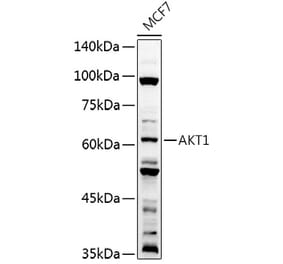
![Western Blot - Anti-AKT1 Antibody [ARC51585] (A308667) - Antibodies.com](https://cdn.antibodies.com/image/catalog/308/A308667_1.jpg?profile=product_alternative)
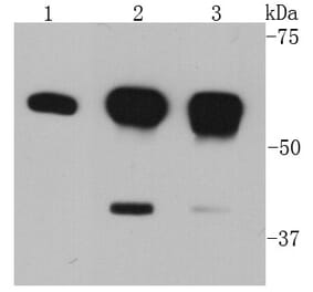
![Western Blot - Anti-AKT1 Antibody [ARC51585] (A309277) - Antibodies.com](https://cdn.antibodies.com/image/catalog/309/A309277_1.jpg?profile=product_alternative)
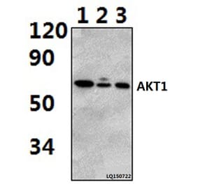
![Western Blot - Anti-AKT1 (phospho Ser473) Antibody [ARC0169] (A80471) - Antibodies.com](https://cdn.antibodies.com/image/catalog/80/A80471_1.jpg?profile=product_alternative)
![Immunohistochemistry - Anti-AKT1 Antibody [AKT1/2784] (A248468) - Antibodies.com](https://cdn.antibodies.com/image/catalog/248/A248468_1.jpg?profile=product_alternative)