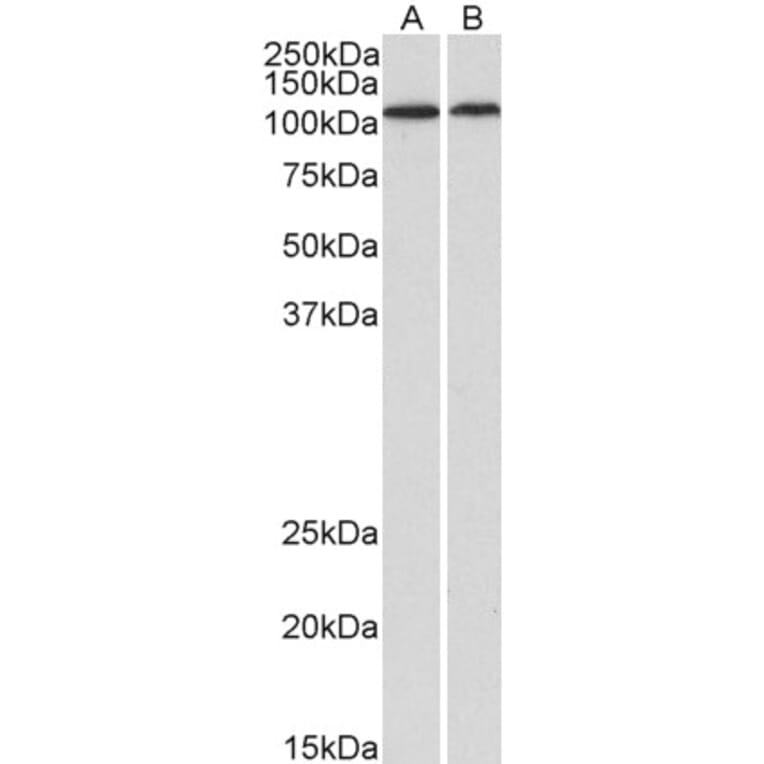
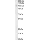
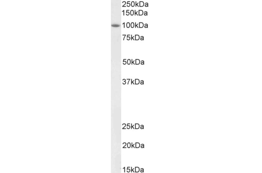
Figure 1: Western Blot - Anti-BMP1/PCP Antibody (A82931)
BMP1/PCP expression in Human Heart (A) and Kidney (B) lysates analyzed by western blot. Cells were lysed in RIPA buffer and 35µg protein was run per lane. Primary antibody incubation was performed with Anti-BMP1/PCP Antibody (A82931) at 1µg/ml and detected by chemiluminescence.
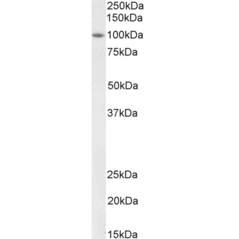
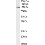
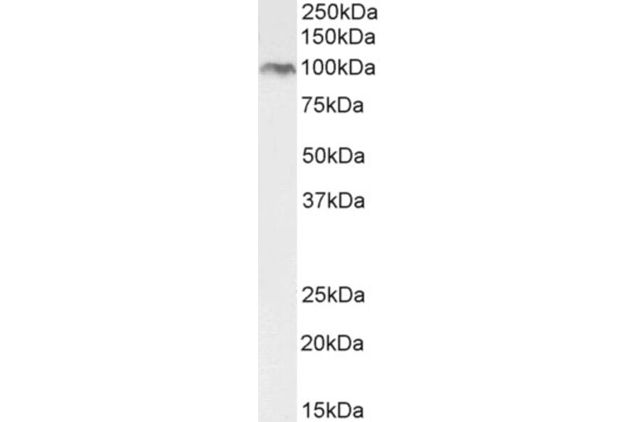
Figure 2: Western Blot - Anti-BMP1/PCP Antibody (A82931)
BMP1/PCP expression in Rat Heart lysate analyzed by western blot. Cells were lysed in RIPA buffer and 35µg protein was run per lane. Primary antibody incubation was performed with Anti-BMP1/PCP Antibody (A82931) at 2µg/ml and detected by chemiluminescence.
Figure 3: Western Blot - Anti-BMP1/PCP Antibody (A82931)
BMP1/PCP expression in NIH3T3 cell lysate analyzed by western blot. Cells were lysed in RIPA buffer and 35µg protein was run per lane. Primary antibody incubation was performed with Anti-BMP1/PCP Antibody (A82931) at 2µg/ml and detected by chemiluminescence.
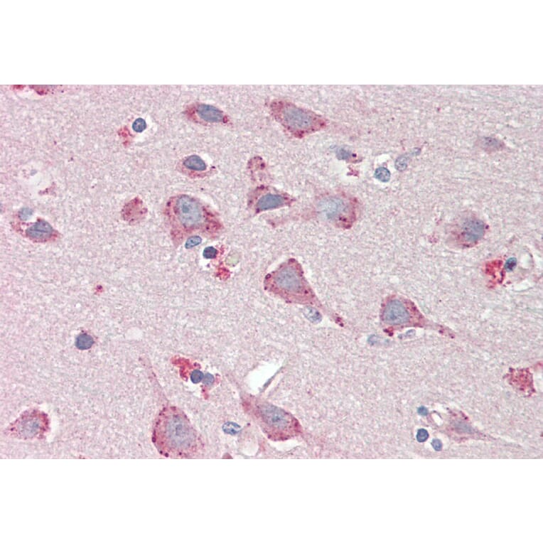
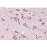
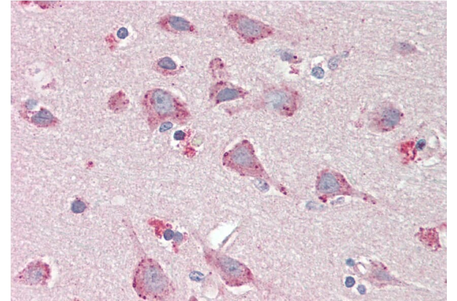
BMP1/PCP expression in Human Cortex analyzed by immunohistochemistry. Tissue was paraffin-embedded, and antigen retrieval was achieved by steaming in citrate buffer, pH 6. Staining was performed with Anti-BMP1/PCP Antibody (A82931) at 5µg/ml and revealed with alkaline phosphatase (AP).
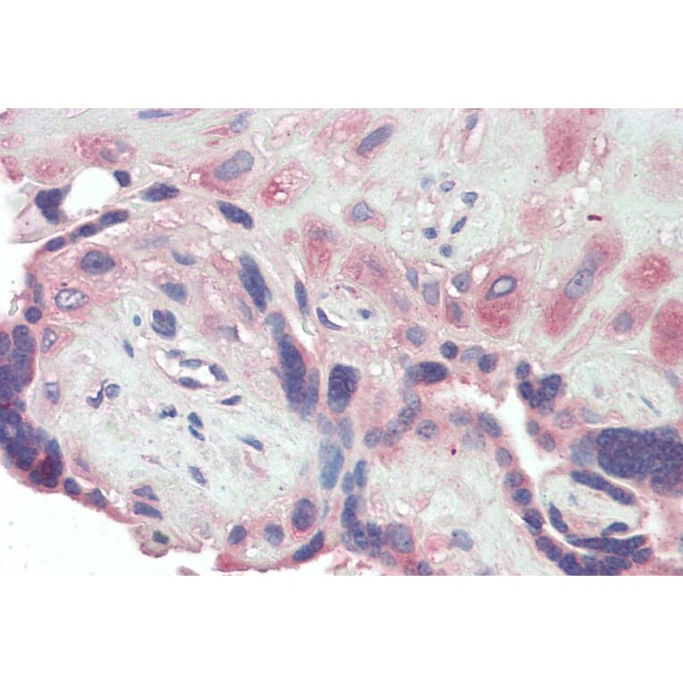
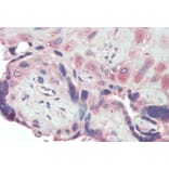
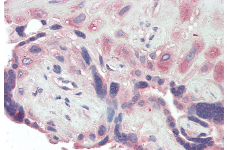
BMP1/PCP expression in Human Placenta analyzed by immunohistochemistry. Tissue was paraffin-embedded, and antigen retrieval was achieved by steaming in citrate buffer, pH 6. Staining was performed with Anti-BMP1/PCP Antibody (A82931) at 5µg/ml and revealed with alkaline phosphatase (AP).