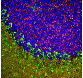
Immunofluorescent analysis of mouse hippocampus section stained with Anti-beta Synuclein Antibody [6A10] (A270558), at a dilution of 1:500, in green, and co-stained with Anti-NF-L Antibody (A270580), at a dilution of 1:5,000, in red. Nuclear DNA is visualised in blue using Hoechst staining. Following transcardial perfusion with 4% paraformaldehyde, the brain was post-fixed for 24 hours, cut to 45 µm, and free-floating sections were stained using the above antibodies. Anti-beta Synuclein Antibody [6A10] (A270558) detects protein concentrated in synaptic regions, while Anti-NF-L Antibody (A270580) labels dendrites and axons of neuronal cells.
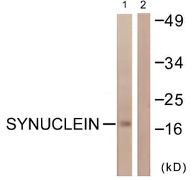
Western Blot - Anti-beta Synuclein Antibody [6A10] (A270558)
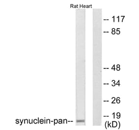
Western blot analysis of different tissue lysates using Anti-beta Synuclein Antibody [6A10] (A270558), at a dilution of 1:1,000, in green. The lanes contain: [Lane 1] protein standard (red), [Lane 2] rat cortex, [Lane 3] rat cerebellum, [Lane 4] mouse cortex, [Lane 5] mouse cerebellum, [Lane 6] cow cortex, and [Lane 7] cow cerebellum. The strong band at about 17 kDa corresponds to beta Synuclein protein.
Immunofluorescent analysis of rat cerebellum section stained with Anti-NF-L Antibody (A270580), at a dilution of 1:5,000, in red, and co-stained with Anti-beta Synuclein Antibody [6A10] (A270558), at a dilution of 1:500, in green. Nuclear DNA is visualised in blue using Hoechst staining. Following transcardial perfusion with 4% paraformaldehyde, the brain was post-fixed for 24 hours, cut to 45 µm, and free-floating sections were stained using the above antibodies. Anti-NF-L Antibody (A270580) labels dendrites and axons of neuronal cells, and Anti-beta Synuclein Antibody [6A10] (A270558) detects protein that is concentrated in synaptic regions.
Immunofluorescent analysis of rat cerebellum section stained with Anti-beta Synuclein Antibody [6A10] (A270558) at a dilution of 1:500 (green) and costained with Anti-Calbindin Antibody (A85359) at a dilution of 1:5,000 (red). Nuclei were stained with Hoechst (blue). Following transcardial perfusion of rat with 4% paraformaldehyde, brain was post fixed for 24 hours, cut to 45 µM, and free-floating sections were stained with above antibodies. The ß-synuclein antibody detects protein concentrated in synaptic regions, and calbindin antibody labels perikarya and dendrites of cerebellar Purkinje cells.
Immunohistochemistry analysis of a 4% PFA fixed paraffin embedded rat hippocampus section with Anti-beta Synuclein Antibody [6A10] (A270558) at a dilution of 1:1,000 detected with DAB (brown) using the Vector Labs ImmPRESS method and reagents with citra buffer retrieval. Counterstained with Hematoxylin (blue). ß-synuclein is concentrated in pre-synaptic regions. Note: this antibody performs well in testing with both 4% PFA and standard NBF fixed tissues but does not stain long term NBF fixed tissue effectively.
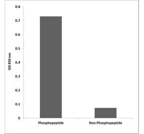
Western Blot - Anti-beta Synuclein Antibody [6A10] (A270558)
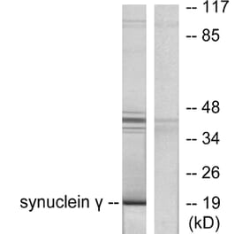
Western blot (left) and Ponceau S stained blot (right) analysis of [Lane 1] protein standard, recombinant full length human [Lane 2] alpha, [Lane 3] beta, and [Lane 4] gamma Synuclein in lanes 2, 3 and 4 respectively. Anti-beta Synuclein Antibody [6A10] (A270558) reacts strongly with beta Synuclein and shows no reaction with the other proteins.


![Immunofluorescence - Anti-beta Synuclein Antibody [6A10] (A270558) - Antibodies.com](https://cdn.antibodies.com/image/catalog/270/A270558_1.jpg?profile=product_top)
![Western Blot - Anti-beta Synuclein Antibody [6A10] (A270558) - Antibodies.com](https://cdn.antibodies.com/image/catalog/270/A270558_2.jpg?profile=product_top)
![Immunofluorescence - Anti-beta Synuclein Antibody [6A10] (A270558) - Antibodies.com](https://cdn.antibodies.com/image/catalog/270/A270558_3.jpg?profile=product_top)
![Immunofluorescence - Anti-beta Synuclein Antibody [6A10] (A270558) - Antibodies.com](https://cdn.antibodies.com/image/catalog/270/A270558_4.jpg?profile=product_top)
![Immunohistochemistry - Anti-beta Synuclein Antibody [6A10] (A270558) - Antibodies.com](https://cdn.antibodies.com/image/catalog/270/A270558_5.jpg?profile=product_top)
![Western Blot - Anti-beta Synuclein Antibody [6A10] (A270558) - Antibodies.com](https://cdn.antibodies.com/image/catalog/270/A270558_6.jpg?profile=product_top)
![Immunofluorescence - Anti-beta Synuclein Antibody [6A10] (A270558) - Antibodies.com](https://cdn.antibodies.com/image/catalog/270/A270558_1.jpg?profile=product_top_thumb)
![Western Blot - Anti-beta Synuclein Antibody [6A10] (A270558) - Antibodies.com](https://cdn.antibodies.com/image/catalog/270/A270558_2.jpg?profile=product_top_thumb)
![Immunofluorescence - Anti-beta Synuclein Antibody [6A10] (A270558) - Antibodies.com](https://cdn.antibodies.com/image/catalog/270/A270558_3.jpg?profile=product_top_thumb)
![Immunofluorescence - Anti-beta Synuclein Antibody [6A10] (A270558) - Antibodies.com](https://cdn.antibodies.com/image/catalog/270/A270558_4.jpg?profile=product_top_thumb)
![Immunohistochemistry - Anti-beta Synuclein Antibody [6A10] (A270558) - Antibodies.com](https://cdn.antibodies.com/image/catalog/270/A270558_5.jpg?profile=product_top_thumb)
![Western Blot - Anti-beta Synuclein Antibody [6A10] (A270558) - Antibodies.com](https://cdn.antibodies.com/image/catalog/270/A270558_6.jpg?profile=product_top_thumb)
![Immunofluorescence - Anti-beta Synuclein Antibody [6A10] (A270558) - Antibodies.com](https://cdn.antibodies.com/image/catalog/270/A270558_1.jpg?profile=product_image)
![Western Blot - Anti-beta Synuclein Antibody [6A10] (A270558) - Antibodies.com](https://cdn.antibodies.com/image/catalog/270/A270558_2.jpg?profile=product_image)
![Immunofluorescence - Anti-beta Synuclein Antibody [6A10] (A270558) - Antibodies.com](https://cdn.antibodies.com/image/catalog/270/A270558_3.jpg?profile=product_image)
![Immunofluorescence - Anti-beta Synuclein Antibody [6A10] (A270558) - Antibodies.com](https://cdn.antibodies.com/image/catalog/270/A270558_4.jpg?profile=product_image)
![Immunohistochemistry - Anti-beta Synuclein Antibody [6A10] (A270558) - Antibodies.com](https://cdn.antibodies.com/image/catalog/270/A270558_5.jpg?profile=product_image)
![Western Blot - Anti-beta Synuclein Antibody [6A10] (A270558) - Antibodies.com](https://cdn.antibodies.com/image/catalog/270/A270558_6.jpg?profile=product_image)