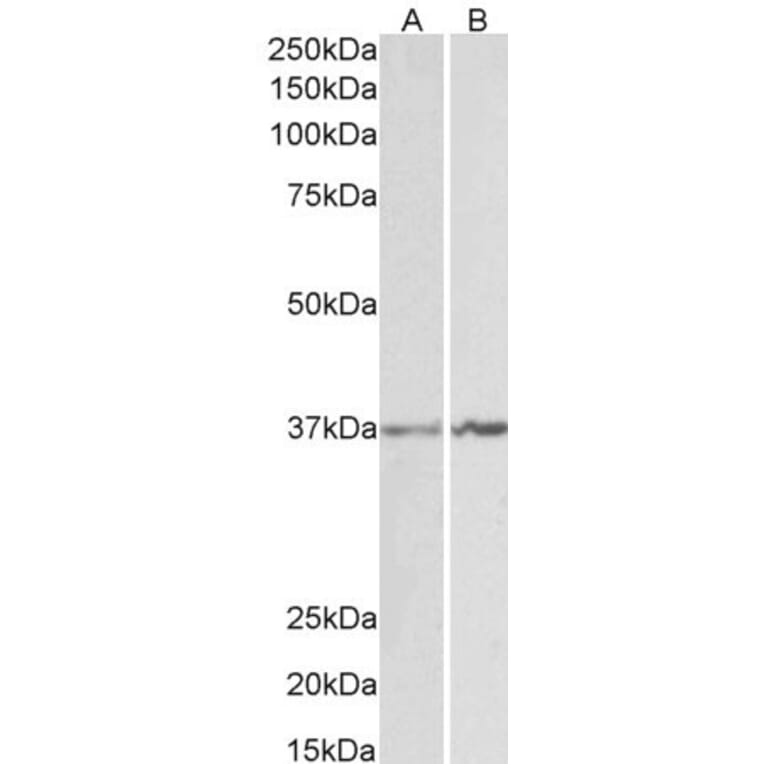
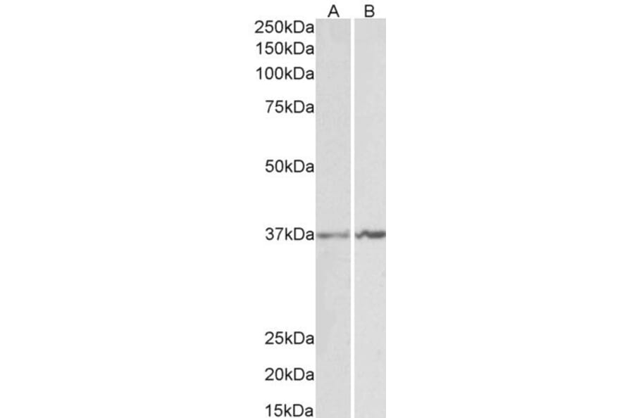
Figure 1: Western Blot - Anti-Annexin A1/ANXA1 Antibody (A84638)
Annexin A1/ANXA1 expression in K562 (A) and NIH3T3 (B) cell lysates analyzed by western blot. Cells were lysed in RIPA buffer and 35µg protein was run per lane. Primary antibody incubation was performed for 1 hour with Anti-Annexin A1/ANXA1 Antibody (A84638) at 1µg/ml and detected by chemiluminescence.
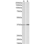
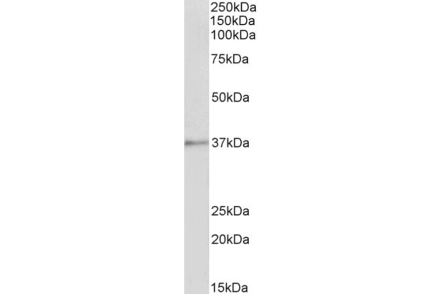
Figure 2: Western Blot - Anti-Annexin A1/ANXA1 Antibody (A84638)
Annexin A1/ANXA1 expression in Rat Spleen lysate analyzed by western blot. Cells were lysed in RIPA buffer and 35µg protein was run per lane. Primary antibody incubation was performed for 1 hour with Anti-Annexin A1/ANXA1 Antibody (A84638) at 1µg/ml and detected by chemiluminescence.
Figure 3: Western Blot - Anti-Annexin A1/ANXA1 Antibody (A84638)
Annexin A1/ANXA1 expression in Pig Spleen lysate analyzed by western blot. Cells were lysed in RIPA buffer and 35µg protein was run per lane. Primary antibody incubation was performed for 1 hour with Anti-Annexin A1/ANXA1 Antibody (A84638) at 1µg/ml and detected by chemiluminescence.
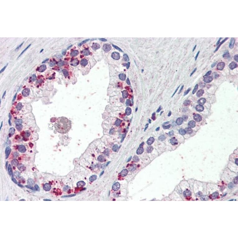
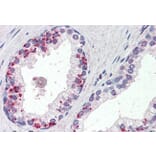
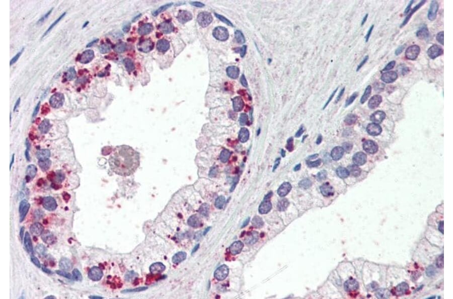
Annexin A1/ANXA1 expression in Human Prostate analyzed by immunohistochemistry. Tissue was paraffin-embedded, and antigen retrieval was achieved by steaming in citrate buffer, pH 6. Staining was performed with Anti-Annexin A1/ANXA1 Antibody (A84638) at 4µg/ml and revealed with alkaline phosphatase (AP). Note this data is from a previous batch and is not on sale.
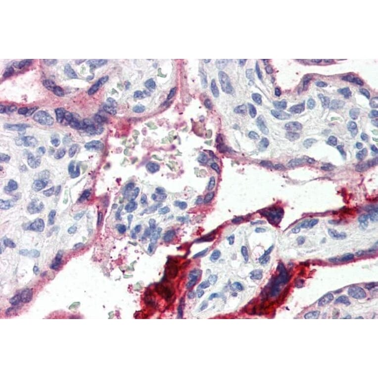
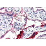
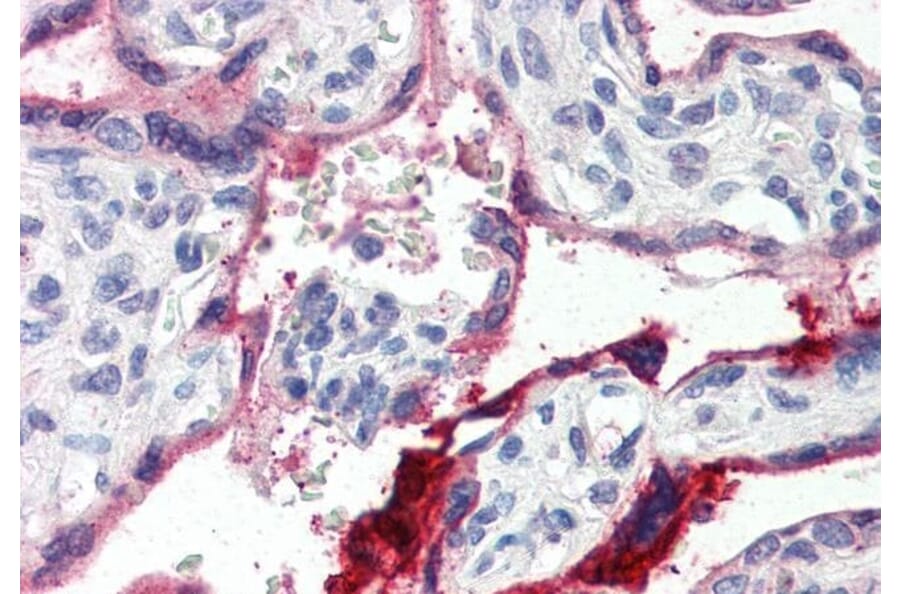
Annexin A1/ANXA1 expression in Human Placenta analyzed by immunohistochemistry. Tissue was paraffin-embedded, and antigen retrieval was achieved by steaming in citrate buffer, pH 6. Staining was performed with Anti-Annexin A1/ANXA1 Antibody (A84638) at 4µg/ml and revealed with alkaline phosphatase (AP). Note this data is from a previous batch and is not on sale.