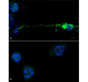
Immunocytochemistry/Immunofluorescence analysis of human iPSC-derived neurons, using Anti-alpha Synuclein (phospho Ser129) Antibody [J18] (A304933), at 1:1,000 for O/N at 4°C. The secondary antibody used was Anti-Rabbit: A488 at 1:1,000 for 1 hour at room temperature. Magnification: 40X. Nuclear stain: Hoechst- 20 min, room temperature (blue). Actin stain: Phalloidin-647- 20 min, room temperature (magenta). 4K cells per well. iPSC neurons: Applied StemCell Catalog # ASE-9321K. A) negative control; no fibrils added to well. B) 7 days after addition of active recombinant human pre-formed fibrils (Type 1). StressMarq catalog # SPR-322. Sonicated before use, 2.5 ug per well.
Western Blot - Anti-alpha Synuclein (phospho Ser129) Antibody [J18] (A304933)
Western blot analysis of mouse brain showing detection of Alpha Synuclein pSer129 protein using Anti-alpha Synuclein (phospho Ser129) Antibody [J18] (A304933) at 1:500 for 2 hours at room temperature with shaking. Lane 1: MW ladder. Lane 2: mouse brain. Load: 15 ug. Block: 5% BSA in TBST. The secondary antibody used was Goat anti-rabbit IgG:HRP at 1:4,000 for 1 hour at room temperature with shaking. Color Development: Chemiluminescent for HRP (Moss) for 5 minutes in room temperature.
Dot Blot - Anti-alpha Synuclein (phospho Ser129) Antibody [J18] (A304933)
Dot blot analysis of alpha synuclein peptide using Anti-alpha Synuclein (phospho Ser129) Antibody [J18] (A304933) at 1:500 for 2 hours at room temperature with shaking. The secondary antibody used was Goat anti-rabbit IgG:HRP at 1:4,000 for 1 hour at room temperature with shaking. Phospho peptide sequence: AYEMP-pS-EEGYQ. Non-phospho peptide sequence: AYEMPSEEGYQ. This sequence is the same for human, mouse, and rat.
Western Blot - Anti-alpha Synuclein (phospho Ser129) Antibody [J18] (A304933)
Western blot analysis of human Alpha Synuclein showing detection of Alpha Synuclein pSer129 protein using Anti-alpha Synuclein (phospho Ser129) Antibody [J18] (A304933) at 1:500 for 2 hours at room temperature with shaking. Lane 6: 15 ug human Parkinson€™s Disease brain lysate.. Block: 5% BSA in TBST. The secondary antibody used was Goat anti-rabbit IgG:HRP at 1:4,000 for 1 hour at room temperature with shaking. Color Development: Chemiluminescent for HRP (Moss) for 5 minutes in room temperature.
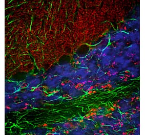
Immunohistochemistry analysis of mouse brain. The Primary Antibody used was Anti-alpha Synuclein (phospho Ser129) Antibody [J18] (A304933) at 8 weeks of age. Mice were unilaterally injected in the dorsal striatum (bregma AP + 0.2 mm, L +/1 2.0 mm, V 3.0 mm) and sacrificed 30 days post-injection.(A) contralateral cortex. (B) ipsilateral cortex. (C) contralateral striatum. (D) ipsilateral striatum.
Publishing research using Anti-alpha Synuclein (phospho Ser129) Antibody [J18] (A304933)? Please let us know so that we can list the citation on this page.
Alternative products to Anti-alpha Synuclein (phospho Ser129) Antibody [J18] (A304933)
Humanized monoclonal antibody to alpha Synuclein Research grade Cinpanemab biosimilar for ELISA, Flow Cytometry, Functional Studies and in vivo Research.
Humanized monoclonal antibody to alpha Synuclein Research grade Cinpanemab biosimilar for ELISA, Flow Cytometry, Functional Studies and in vivo Research.


![Immunocytochemistry/Immunofluorescence - Anti-alpha Synuclein (phospho Ser129) Antibody [J18] (A304933) - Antibodies.com](https://cdn.antibodies.com/image/catalog/304/A304933_1.png?profile=product_top)
![Western Blot - Anti-alpha Synuclein (phospho Ser129) Antibody [J18] (A304933) - Antibodies.com](https://cdn.antibodies.com/image/catalog/304/A304933_2.png?profile=product_top)
![Dot Blot - Anti-alpha Synuclein (phospho Ser129) Antibody [J18] (A304933) - Antibodies.com](https://cdn.antibodies.com/image/catalog/304/A304933_3.png?profile=product_top)
![Western Blot - Anti-alpha Synuclein (phospho Ser129) Antibody [J18] (A304933) - Antibodies.com](https://cdn.antibodies.com/image/catalog/304/A304933_4.png?profile=product_top)
![Immunohistochemistry - Anti-alpha Synuclein (phospho Ser129) Antibody [J18] (A304933) - Antibodies.com](https://cdn.antibodies.com/image/catalog/304/A304933_5.png?profile=product_top)
![Immunohistochemistry - Anti-alpha Synuclein (phospho Ser129) Antibody [J18] (A304933) - Antibodies.com](https://cdn.antibodies.com/image/catalog/304/A304933_6.png?profile=product_top)
![Immunocytochemistry/Immunofluorescence - Anti-alpha Synuclein (phospho Ser129) Antibody [J18] (A304933) - Antibodies.com](https://cdn.antibodies.com/image/catalog/304/A304933_7.png?profile=product_top)
![Immunocytochemistry/Immunofluorescence - Anti-alpha Synuclein (phospho Ser129) Antibody [J18] (A304933) - Antibodies.com](https://cdn.antibodies.com/image/catalog/304/A304933_1.png?profile=product_top_thumb)
![Western Blot - Anti-alpha Synuclein (phospho Ser129) Antibody [J18] (A304933) - Antibodies.com](https://cdn.antibodies.com/image/catalog/304/A304933_2.png?profile=product_top_thumb)
![Dot Blot - Anti-alpha Synuclein (phospho Ser129) Antibody [J18] (A304933) - Antibodies.com](https://cdn.antibodies.com/image/catalog/304/A304933_3.png?profile=product_top_thumb)
![Western Blot - Anti-alpha Synuclein (phospho Ser129) Antibody [J18] (A304933) - Antibodies.com](https://cdn.antibodies.com/image/catalog/304/A304933_4.png?profile=product_top_thumb)
![Immunohistochemistry - Anti-alpha Synuclein (phospho Ser129) Antibody [J18] (A304933) - Antibodies.com](https://cdn.antibodies.com/image/catalog/304/A304933_5.png?profile=product_top_thumb)
![Immunohistochemistry - Anti-alpha Synuclein (phospho Ser129) Antibody [J18] (A304933) - Antibodies.com](https://cdn.antibodies.com/image/catalog/304/A304933_6.png?profile=product_top_thumb)
![Immunocytochemistry/Immunofluorescence - Anti-alpha Synuclein (phospho Ser129) Antibody [J18] (A304933) - Antibodies.com](https://cdn.antibodies.com/image/catalog/304/A304933_7.png?profile=product_top_thumb)
![Immunocytochemistry/Immunofluorescence - Anti-alpha Synuclein (phospho Ser129) Antibody [J18] (A304933) - Antibodies.com](https://cdn.antibodies.com/image/catalog/304/A304933_1.png?profile=product_image)
![Western Blot - Anti-alpha Synuclein (phospho Ser129) Antibody [J18] (A304933) - Antibodies.com](https://cdn.antibodies.com/image/catalog/304/A304933_2.png?profile=product_image)
![Dot Blot - Anti-alpha Synuclein (phospho Ser129) Antibody [J18] (A304933) - Antibodies.com](https://cdn.antibodies.com/image/catalog/304/A304933_3.png?profile=product_image)
![Western Blot - Anti-alpha Synuclein (phospho Ser129) Antibody [J18] (A304933) - Antibodies.com](https://cdn.antibodies.com/image/catalog/304/A304933_4.png?profile=product_image)
![Immunohistochemistry - Anti-alpha Synuclein (phospho Ser129) Antibody [J18] (A304933) - Antibodies.com](https://cdn.antibodies.com/image/catalog/304/A304933_5.png?profile=product_image)
![Immunohistochemistry - Anti-alpha Synuclein (phospho Ser129) Antibody [J18] (A304933) - Antibodies.com](https://cdn.antibodies.com/image/catalog/304/A304933_6.png?profile=product_image)
![Immunocytochemistry/Immunofluorescence - Anti-alpha Synuclein (phospho Ser129) Antibody [J18] (A304933) - Antibodies.com](https://cdn.antibodies.com/image/catalog/304/A304933_7.png?profile=product_image)
![Immunofluorescence - Anti-alpha Synuclein Antibody [2A7] (A85290) - Antibodies.com](https://cdn.antibodies.com/image/catalog/85/A85290_1.jpg?profile=product_alternative)
![Western Blot - Anti-alpha Synuclein Antibody [3C11] (A304959) - Antibodies.com](https://cdn.antibodies.com/image/catalog/304/A304959_1.png?profile=product_alternative)
![Immunocytochemistry/Immunofluorescence - Anti-alpha Synuclein Antibody [4F1] (A304962) - Antibodies.com](https://cdn.antibodies.com/image/catalog/304/A304962_1.png?profile=product_alternative)
![Immunocytochemistry/Immunofluorescence - Anti-alpha Synuclein Antibody [3F8] (A304961) - Antibodies.com](https://cdn.antibodies.com/image/catalog/304/A304961_1.png?profile=product_alternative)
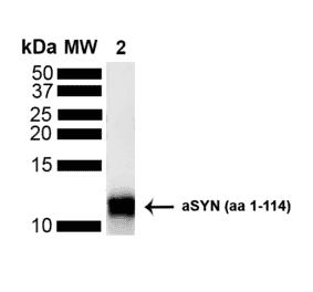
![SDS-PAGE - Anti-alpha Synuclein Antibody [MEDI1341] Biosimilar - BSA and Azide free (A339878) - Antibodies.com](https://cdn.antibodies.com/image/catalog/339/A339878_1.jpg?profile=product_alternative)
![Western Blot - Anti-alpha Synuclein Antibody [10H7] (A304960) - Antibodies.com](https://cdn.antibodies.com/image/catalog/304/A304960_1.png?profile=product_alternative)