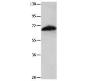
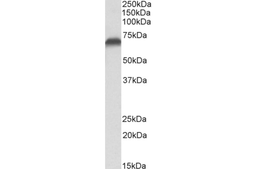
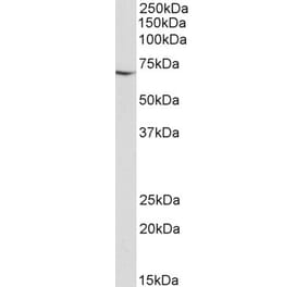
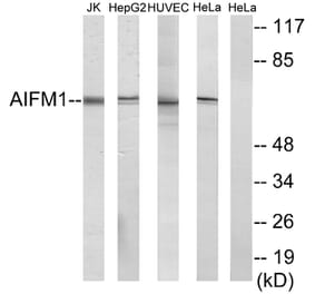
AIF expression in Jurkat cell lysate analyzed by western blot. Cells were lysed in RIPA buffer and 35µg protein was run per lane. Primary antibody incubation was performed with Anti-AIF Antibody (A82698) at 0.1µg/ml and detected by chemiluminescence.
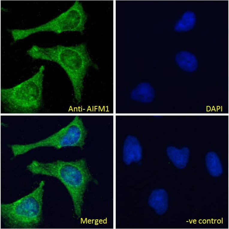
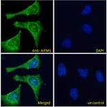
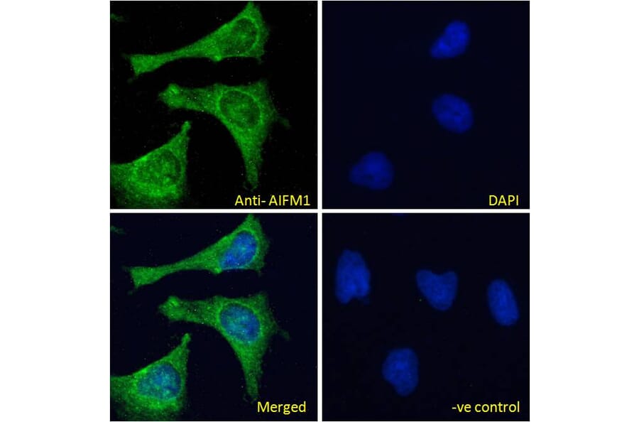
AIF expression in HeLa cells analyzed by immunofluorescence. Cells were permeabilized with 0.15% Triton. Staining was performed with Anti-AIF Antibody (A82698) at 10µg/ml for 1 hour and Alexa Fluor 488 secondary antibody at 4µg/ml. Mitochondrial staining shown and nuclei were stained with DAPI (blue). Negative control: Goat IgG Isotype Control at 10µg/ml followed by Alexa Fluor 488 secondary antibody at 4µg/ml.
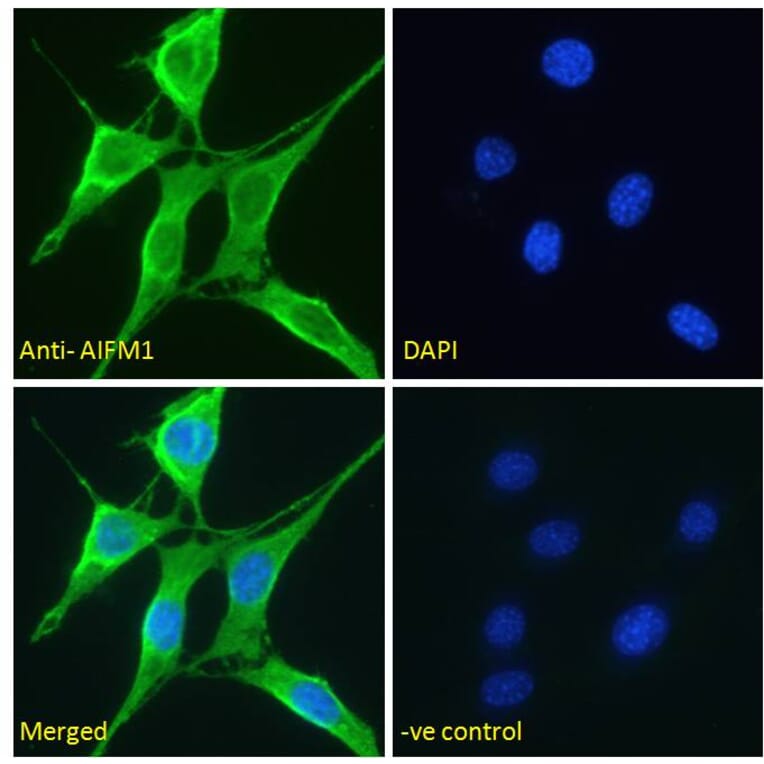
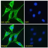
AIF expression in NIH3T3 cells analyzed by immunofluorescence. Cells were permeabilized with 0.15% Triton. Staining was performed with Anti-AIF Antibody (A82698) at 5µg/ml for 1 hour and Alexa Fluor 488 secondary antibody at 2µg/ml. Mitochondrial staining shown and nuclei were stained with DAPI (blue). Negative control: Goat IgG Isotype Control at 5µg/ml followed by Alexa Fluor 488 secondary antibody at 2µg/ml.
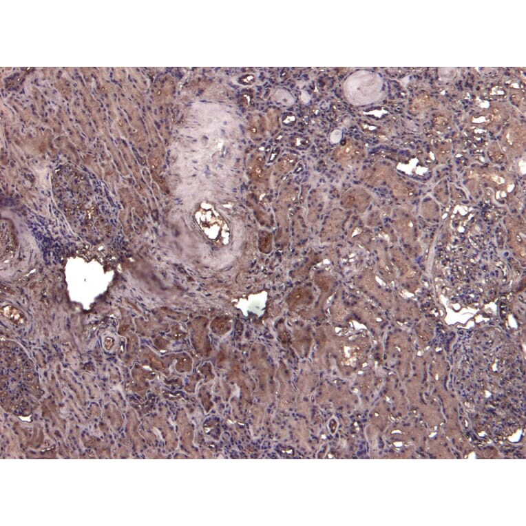
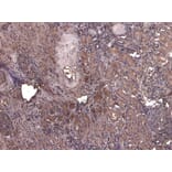
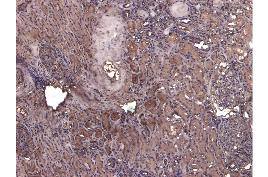
AIF expression in Human Kidney analyzed by immunohistochemistry. Tissue was paraffin-embedded, and antigen retrieval was achieved by heating in citrate buffer, pH 6. Staining was performed with Anti-AIF Antibody (A82698) at 5µg/ml and revealed with horseradish peroxidase (HRP).
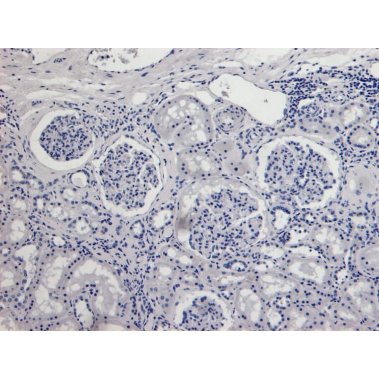
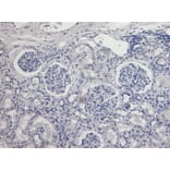
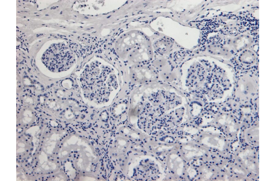
Negative control for AIF expression in Human Kidney analyzed by immunohistochemistry. Tissue was paraffin-embedded, and staining procedure was performed in the absence of primary antibody.


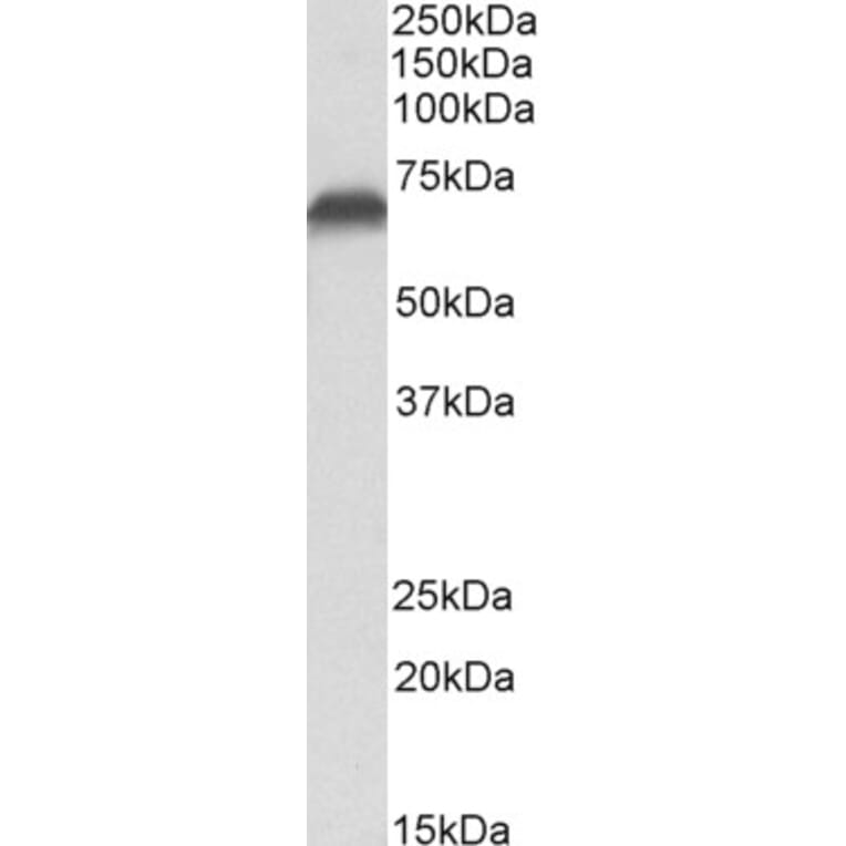


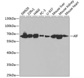
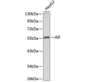
![Western Blot - Anti-AIF Antibody [ARC0015] (A308543) - Antibodies.com](https://cdn.antibodies.com/image/catalog/308/A308543_1.jpg?profile=product_alternative)